. 2011 Mar;134(Pt 3):903-12.
doi: 10.1093/brain/awq355. Epub 2010 Dec 13.

The anatomy underlying acute versus chronic spatial neglect: a longitudinal study

Affiliations

The anatomy underlying acute versus chronic spatial neglect: a longitudinal study

Hans-Otto Karnath et al. Brain. 2011 Mar.

Abstract

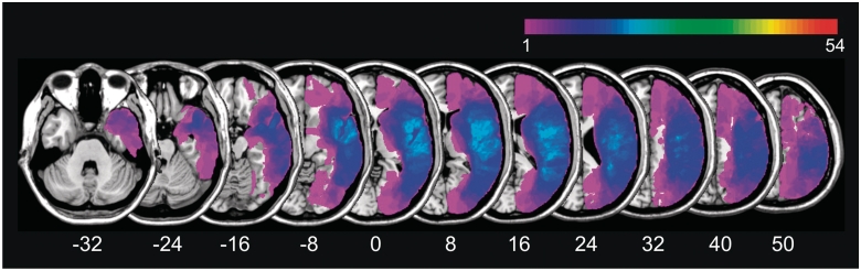
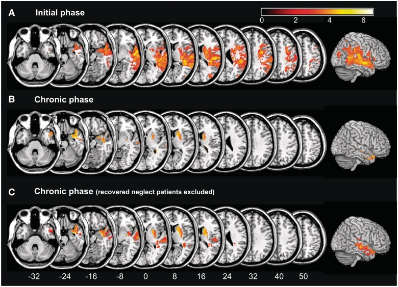
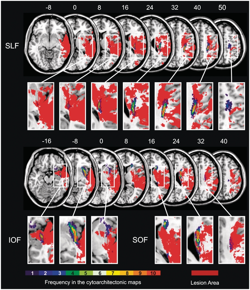
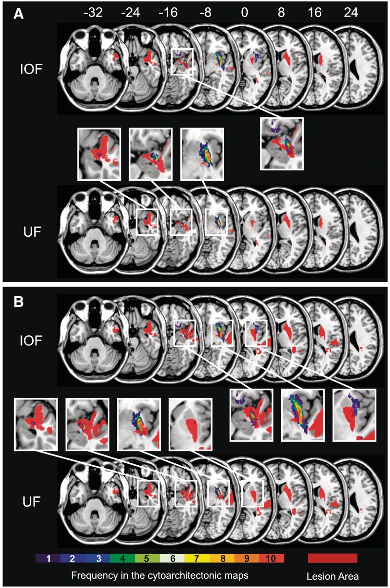
Our aim was to examine how brain imaging in the initial phase of a stroke could predict both acute/subacute as well as chronic spatial neglect. We present the first voxel-wise longitudinal lesion-behaviour mapping study, examining acute/subacute as well as chronic performance in the same individuals. Acute brain imaging (acquired on average 6.2 days post-injury) was used to evaluate neglect symptoms at the initial (mean 12.4 days post-stroke) and the chronic (mean 491 days) phase of the stroke. Chronic neglect was found in about one-third of the patients with acute neglect. Analysis suggests that lesion of the superior and middle temporal gyri predict both acute/subacute as well as chronic neglect. At the subcortical level, the basal ganglia as well as the inferior occipitofrontal fasciculus/extreme capsule appear to play a significant role for both acute/subacute as well as chronic neglect. Beyond, the uncinate fasciculus was critically related to the emergence of chronic spatial neglect. We infer that individuals who experience spatial neglect in the initial phase of the stroke yet do not have injury to these cortical and subcortical structures are likely to recover, and thus have a favourable prognosis.


Figures

Figure 1
Figure 1
Simple overlap of all 54 stroke patients with right-hemisphere brain lesions.
Figure 2
Figure 2
Statistical voxel-wise lesion-behaviour mapping (VLBM) analyses using the t-test statistic for the continuous variable ‘neglect severity’. Presented are all voxels that survived a 1% false discovery rate cut-off threshold. (A) Analysis of all 54 patients with right hemisphere damage based on their neglect severity scores measured in the initial phase of the stroke, on average 12.4 days after stroke onset. Injury to highlighted regions predicts acute/subacute neglect. (B) Analysis of all 54 patients with right hemisphere damage based on their neglect severity scores measured in the chronic phase of the stroke, on average 490.8 days after stroke onset. Injury to highlighted regions predicts chronic neglect, regardless of acute behaviour. (C) Analysis of the neglect severity scores measured in the chronic phase of the stroke as in (B), but now carried out on a subsample (n = 38) of the patients with right hemisphere damage. This subsample no longer included those patients that showed spatial neglect in the initial phase of the stroke but had recovered by the time of the second examination. Montreal Neurological Institute coordinates of each transverse section are given.
Figure 3
Figure 3
White matter fibre tract analysis in the initial phase of the stroke. Overlap of the statistical lesion map from Fig. 2A (in homogenous orange colour) with the probabilistic, cytoarchitectonic maps of the white matter fibre tracts from the Jülich atlas (right hemisphere only). The colour coding of the atlas from 1 (dark blue, observed in one post-mortem brain) to 10 (red, overlap in all 10 post-mortem brains) represents the absolute frequency for which, in each voxel of the atlas, a respective fibre tract was present. The overlap is illustrated for the superior longitudinal fasciculus (SLF), inferior occipitofrontal fasciculus (IOF) and superior occipitofrontal fasciculus (SOF).
Figure 4
Figure 4
White matter fibre tract analysis in the chronic phase of the stroke. Overlap of the statistical lesion map from Fig. 2B and C (in homogenous orange colour) with the probabilistic, cytoarchitectonic maps of the white matter fibre tracts from the Jülich atlas (right hemisphere only). Data deriving from the analysis of all 54 patients with right hemisphere damage (A) and of the subsample of this group that no longer included those patients that showed spatial neglect in the initial phase of the stroke but had recovered by the time of the second examination (B). The colour coding of the atlas from 1 (dark blue, observed in one post-mortem brain) to 10 (red, overlap in all 10 post-mortem brains) represents the absolute frequency for which, in each voxel of the atlas, a respective fibre tract was present. The overlap is illustrated for the inferior occipitofrontal fasciculus (IOF) and uncinate fasciculus (UF).

Comment in

References

    1. Bates E, Wilson SM, Saygin AP, Dick F, Sereno MI, Knight RT, et al. Voxel-based lesion-symptom mapping. Nat Neurosci. 2003;6:448–50. - PubMed
    1. Becker E, Karnath H-O. Neuroimaging of eye position reveals spatial neglect. Brain. 2010;133:909–14. - PubMed
    1. Black S, Ebert P, Leibovitch F, Szalai JP, Blair N. Recovery in hemispatial neglect. Neurology. 1995;45(Suppl. 4):A178. - PubMed
    1. Brett M, Leff AP, Rorden C, Ashburner J. Spatial normalisation of brain images with focal lesions using cost function masking. Neuroimage. 2001;14:486–500. - PubMed
    1. Bürgel U, Amunts K, Hoemke L, Gilsbach JM, Zilles K. White matter fiber tracts of the human brain: three-dimensional mapping at microscopic resolution, topography and intersubject variability. Neuroimage. 2006;29:1092–105. - PubMed

Publication types