. 2011 Feb 25;286(8):6697-706.
doi: 10.1074/jbc.M110.183558. Epub 2010 Dec 14.

Ca2+/calmodulin-dependent protein kinase II inhibitors disrupt AKAP79-dependent PKC signaling to GluA1 AMPA receptors

Affiliations

Ca2+/calmodulin-dependent protein kinase II inhibitors disrupt AKAP79-dependent PKC signaling to GluA1 AMPA receptors

Ian M Brooks et al. J Biol Chem. .

Abstract

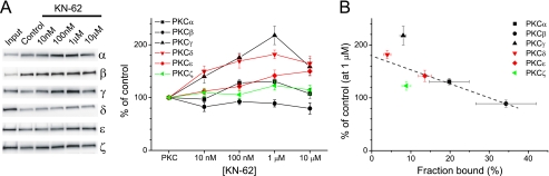
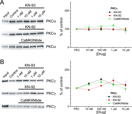
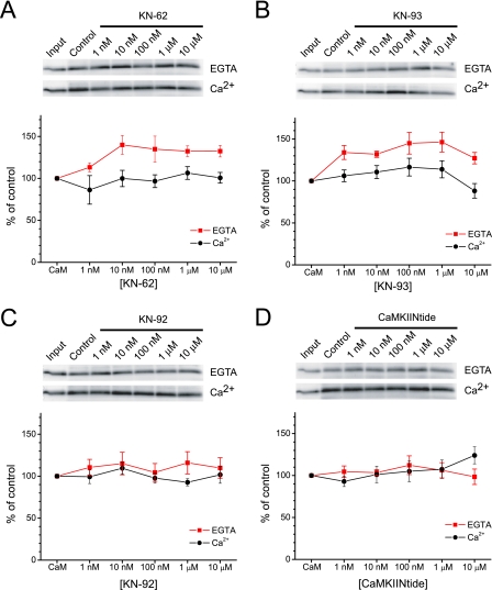
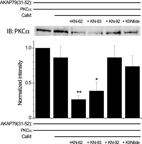
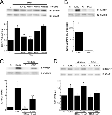
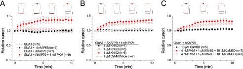
GluA1 (formerly GluR1) AMPA receptor subunit phosphorylation at Ser-831 is an early biochemical marker for long-term potentiation and learning. This site is a substrate for Ca(2+)/calmodulin (CaM)-dependent protein kinase II (CaMKII) and protein kinase C (PKC). By directing PKC to GluA1, A-kinase anchoring protein 79 (AKAP79) facilitates Ser-831 phosphorylation and makes PKC a more potent regulator of GluA1 than CaMKII. PKC and CaM bind to residues 31-52 of AKAP79 in a competitive manner. Here, we demonstrate that common CaMKII inhibitors alter PKC and CaM interactions with AKAP79(31-52). Most notably, the classical CaMKII inhibitors KN-93 and KN-62 potently enhanced the association of CaM to AKAP79(31-52) in the absence (apoCaM) but not the presence of Ca(2+). In contrast, apoCaM association to AKAP79(31-52) was unaffected by the control compound KN-92 or a mechanistically distinct CaMKII inhibitor (CaMKIINtide). In vitro studies demonstrated that KN-62 and KN-93, but not the other compounds, led to apoCaM-dependent displacement of PKC from AKAP79(31-52). In the absence of CaMKII activation, complementary cellular studies revealed that KN-62 and KN-93, but not KN-92 or CaMKIINtide, inhibited PKC-mediated phosphorylation of GluA1 in hippocampal neurons as well as AKAP79-dependent PKC-mediated augmentation of recombinant GluA1 currents. Buffering cellular CaM attenuated the ability of KN-62 and KN-93 to inhibit AKAP79-anchored PKC regulation of GluA1. Therefore, by favoring apoCaM binding to AKAP79, KN-62 and KN-93 derail the ability of AKAP79 to efficiently recruit PKC for regulation of GluA1. Thus, AKAP79 endows PKC with a pharmacological profile that overlaps with CaMKII.


Figures

FIGURE 1.
FIGURE 1.
KN-62 affects AKAP79-PKC interactions in isoform-dependent manner. AKAP79(31–52) was incubated with PKC isoforms (200 ng; Input lane, 25 ng) and KN-62 as indicated. A, left, representative blots showing the effect of KN-62 on PKC isoform binding AKAP79(31–52). Right, graphical summary of data normalized to their respective controls. Statistical significance for each group is as follows: PKCα: n = 6; 100 nm and 1 μm, p < 0.05; PKCβ: n = 4–5; not significant; PKCγ: n = 5–6; 100 nm and 1 μm, p < 0.01; 10 μm, p < 0.05; PKCδ: n = 4–5; 10 nm–10 μm, p < 0.01; PKCϵ: n = 7–8; 1–10 μm, p < 0.01; and PKCζ: n = 4–5; not significant. B, percentage of control binding to AKAP79(31–52) in response to KN-62 (1 μm) is plotted versus the fraction of input bound for each PKC isoform. Percentages are calculated as the amount of PKC recovered in control lanes normalized to the input lane. Black, red, and green symbols reflect typical, novel, and atypical PKC isoforms, respectively. The dashed line represents the best fit through the data determined by linear regression analysis (r = 0.85). All graphs depict mean ± S.E.
FIGURE 2.
FIGURE 2.
Differential effects of CaMKII inhibitors on PKCα and PKCγ. AKAP79(31–52) was incubated with PKCα and KN-93 (n = 3–4), KN-92 (n = 3–4), or CaMKIINtide (n = 8) as indicated (A) or PKCγ and KN-93 (n = 4–5; 100 nm, p < 0.01), KN-92 (n = 3; 1 μm, p < 0.05), or CaMKIINtide (n = 3–4; 100 nm, p < 0.05) (B). Left panels, representative blots for each condition. Right panels, summary data normalized to respective controls. All graphs depict mean ± S.E.
FIGURE 3.
FIGURE 3.
Classical CaMKII inhibitors enhance Ca2+-independent binding of CaM to AKAP79. AKAP79(31–52) was incubated with CaM (1 μm; Input lane, 50 ng) and KN-62 at the indicated concentrations in the absence or presence of Ca2+. A, top, representative blots of CaM binding to AKAP79(31–52) in 1 mm EGTA (upper) or 100 μm Ca2+ (lower). Bottom, summary data normalized to respective controls (n = 3–6; for EGTA: 10 nm–10 μm KN-62, p < 0.05). B, same as A but with KN-93 (n = 5–7; for EGTA: 1 nm, 100 nm, and 1 μm, p < 0.01; 10 nm, p < 0.05). C and D, same as above except with KN-92 (n = 6) and CaMKIINtide (n = 5–6), respectively. All graphs depict mean ± S.E.
FIGURE 4.
FIGURE 4.
Classical CaMKII inhibitors induce apoCaM-mediated displacement of PKC from AKAP79. AKAP(31–52) was incubated with PKCα (200 ng) in the absence or presence of apoCaM (10 μm) and various CaMKII reagents (each 1 μm). Top, representative Western blot of PKCα binding to AKAP79(31–52) in the absence or presence of apoCaM and the indicated CaMKII reagents. Bottom, graph summarizing the data (mean ± S.E.) from multiple experiments (n = 6). For each experiment, the data were normalized to the amount of PKC bound to AKAP79(31–52) alone. *, p < 0.05; **, p < 0.01 compared with PKCα + AKAP79(31–52) + apoCaM. KIINtide, CaMKIINtide; IB, immunoblot.
FIGURE 5.
FIGURE 5.
Classical CaMKII inhibitors block PKC phosphorylation of GluA1 in neurons. A, hippocampal neurons were untreated (i.e. control (C)) or pretreated with CaMKII reagents (10 μm each) as indicated prior to PMA application (1 μm; 3 min) to activate PKC. Top, representative blots of Ser-831 phosphorylation (S831P) visualized using a phosphospecific antibody (upper panel) or to total GluA1 (lower panel). Bottom, summary graph of the ratio of Ser-831 phosphorylation/GluR1 signals normalized to control (n = 8–13; *, p < 0.01 compared with control; **, p < 0.01 compared with PMA). B, neurons were untreated (C) or stimulated with ionomycin (IONO; 10 μm; 3 min) or PMA (1 μm; 3 min). Top, representative blots demonstrating increase in CaMKII phosphorylation at Thr-286 (T286P) in response to ionomycin but not to PMA (upper panel). Blots were reprobed with an antibody directed against CaMKII (lower panel). Bottom, summary graph of the ratio of phosphorylation at Thr-286/CaMKII signals as percentage of control (n = 3; *, p < 0.05 compared with control). C, neurons were untreated (C) or stimulated with ionomycin (10 μm; 3 min) with or without CaMKIINtide (KIINtide; 10 μm; 15 min) pretreatment. Top, representative blots demonstrating CaMKIINtide suppression of basal and ionomycin-induced phosphorylation of CaMKII at Thr-286 (T286P) (upper panel). Blots were reprobed with an antibody directed against CaMKII (lower panel). Bottom, summary graph of the ratio of phosphorylation of CaMKII at Thr-286/CaMKII signals normalized to control (n = 7; *, p < 0.01 compared with control; **, p < 0.05 compared with the corresponding condition in the absence of CaMKIINtide). D, neurons were untreated (C) or stimulated with ionomycin (10 μm; 3 min) with or without CaMKIINtide (KIINtide; 10 μm; 15 min) or BIS-I (0.5 μm; 15 min) pretreatment. Top, representative blots demonstrating that the PKC inhibition by BIS-I but not CaMKII inhibition by CaMKIINtide suppressed the ionomycin-induced phosphorylation of GluA1 at Ser-831 (S831P) (upper panel). Blots were reprobed with an antibody directed against GluR1 (lower panel). Bottom, summary graph of the ratio of phosphorylation of GluA1 at Ser-831/GluA1 signals normalized to control (n = 6; *, p < 0.01 compared with their respective controls). IB, immunoblot. All graphs depict mean ± S.E.
FIGURE 6.
FIGURE 6.
Classical CaMKII inhibitors prevent AKAP79-anchored PKC regulation of GluA1. A, HEK cells were transfected with GluA1 ± AKAP79. A summary time course of GluA1 receptor currents is shown, demonstrating that AKAP79 facilitates PKC regulation of GluA1 (GluA1 AKAP79 versus GluA1 + AKAP79 + PKM, p < 0.05). PKM (4 nm) was included in the patch pipette as indicated. All data are expressed as mean ± S.E. The number of observations for each condition is indicated. Insets, representative glutamate-evoked (500-ms) current traces from the first (black) and final (red) sweep (10 min) for each condition. Vertical scale bars equal 500 pA. B, KN-62 and KN-93, but not KN-92 or CaMKIINtide, inhibit AKAP79-anchored PKC regulation of GluR1 (KN-62 and KN-93 both p < 0.05 compared with GluA1 + AKAP79 + PKM from A). Cells were transfected with GluA1 + AKAP79. Patch pipettes contained PKM. Cells were pretreated with KN-62, KN-93, KN-92, or CaMKIINtide and recorded in the continued presence of these reagents. Data are depicted as in A except that vertical scale bars equal 1 nA. C, buffering CaM by infusion of the CaMBD (10 μm) restores AKAP79-anchored PKC-mediated up-regulation of GluA1 receptor currents in the presence of KN-62 and KN-93. Cells were transfected with GluA1 + AKAP79. Cells were infused with the CaMBD alone or the CaMBD + PKM in the presence of KN-62 or KN-93. Data are depicted as in A except that vertical scale bars equal 1 nA. The CaMBD alone did not modify the stability of GluR1 receptor currents (compare with GluA1 + AKAP79 in A). However, infusion of the CaMBD prevented KN-62- and KN-93-mediated inhibition of AKAP79-anchored PKC regulation of GluA1 (both p < 0.05 compared with the corresponding treatments in B).

Similar articles

Cited by

References

    1. Davies S. P., Reddy H., Caivano M., Cohen P. (2000) Biochem. J. 351, 95–105 - PMC - PubMed
    1. Tokumitsu H., Chijiwa T., Hagiwara M., Mizutani A., Terasawa M., Hidaka H. (1990) J. Biol. Chem. 265, 4315–4320 - PubMed
    1. Sumi M., Kiuchi K., Ishikawa T., Ishii A., Hagiwara M., Nagatsu T., Hidaka H. (1991) Biochem. Biophys. Res. Commun. 181, 968–975 - PubMed
    1. Gargett C. E., Wiley J. S. (1997) Br. J. Pharmacol. 120, 1483–1490 - PMC - PubMed
    1. Humphreys B. D., Virginio C., Surprenant A., Rice J., Dubyak G. R. (1998) Mol. Pharmacol. 54, 22–32 - PubMed

Publication types

MeSH terms

Substances