. 2011 Feb 18;286(7):5546-57.
doi: 10.1074/jbc.M110.183699. Epub 2010 Dec 14.

Ursolic acid, a pentacyclin triterpene, potentiates TRAIL-induced apoptosis through p53-independent up-regulation of death receptors: evidence for the role of reactive oxygen species and JNK

Affiliations

Ursolic acid, a pentacyclin triterpene, potentiates TRAIL-induced apoptosis through p53-independent up-regulation of death receptors: evidence for the role of reactive oxygen species and JNK

Sahdeo Prasad et al. J Biol Chem. .

Retraction in

Abstract

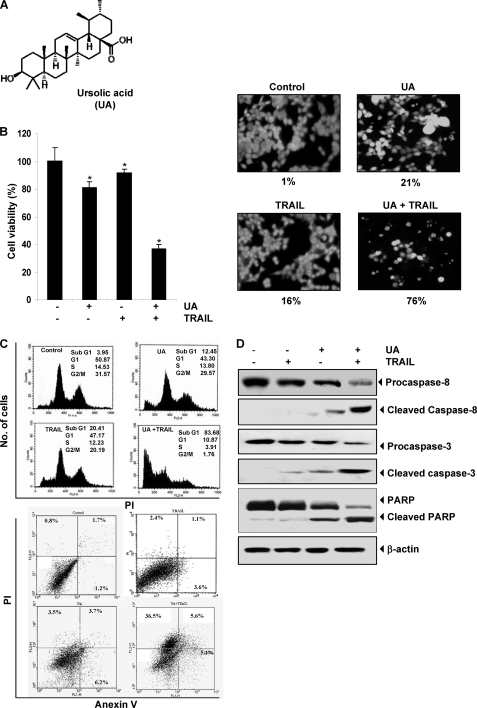
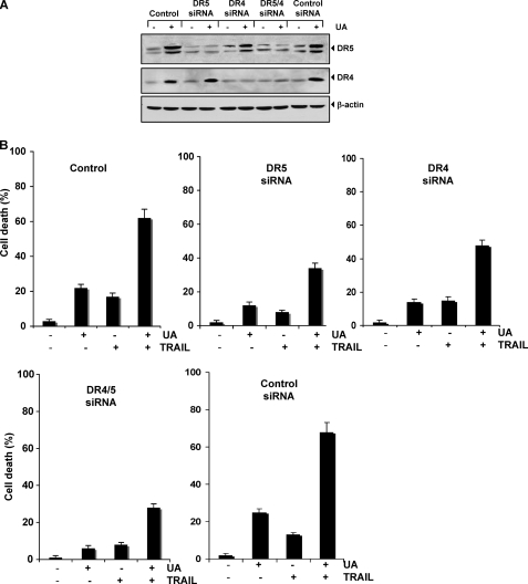
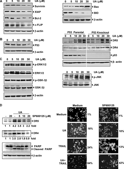
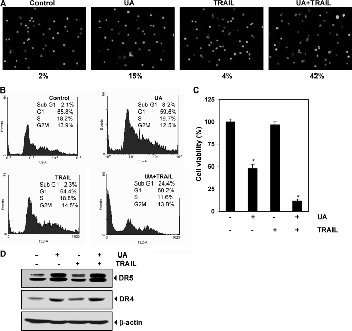
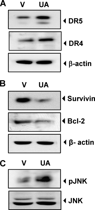
Discovery of the molecular targets of traditional medicine and its chemical footprints can validate the use of such medicine. In the present report, we investigated the effect of ursolic acid (UA), a pentacyclic triterpenoid found in rosemary and holy basil, on apoptosis induced by TRAIL. We found that UA potentiated TRAIL-induced apoptosis in cancer cells. In addition, UA also sensitized TRAIL-resistant cancer cells to the cytokine. When we investigated the mechanism, we found that UA down-regulated cell survival proteins and induced the cell surface expression of both TRAIL receptors, death receptors 4 and 5 (DR4 and -5). Induction of receptors by UA occurred independently of cell type. Gene silencing of either receptor by small interfering RNA reduced the apoptosis induced by UA and the effect of TRAIL. In addition, UA also decreased the expression of decoy receptor 2 (DcR2) but not DcR1. Induction of DRs was independent of p53 because UA induced DR4 and DR5 in HCT116 p53(-/-) cells. Induction of DRs, however, was dependent on JNK because UA induced JNK, and its pharmacologic inhibition abolished the induction of the receptors. The down-regulation of survival proteins and up-regulation of the DRs required reactive oxygen species (ROS) because UA induced ROS, and its quenching abolished the effect of the terpene. Also, potentiation of TRAIL-induced apoptosis by UA was significantly reduced by both ROS quenchers and JNK inhibitor. In addition, UA was also found to induce the expression of DRs, down-regulate cell survival proteins, and activate JNK in orthotopically implanted human colorectal cancer in a nude mouse model. Overall, our results showed that UA potentiates TRAIL-induced apoptosis through activation of ROS and JNK-mediated up-regulation of DRs and down-regulation of DcR2 and cell survival proteins.


Figures

FIGURE 1.
FIGURE 1.
UA potentiates TRAIL-induced apoptosis of HCT116 cells. A, chemical structure of UA. B, cells were pretreated with 20 μm UA for 12 h. The medium was removed, and the cells were then exposed to TRAIL (25 ng/ml) for 24 h. Cell viability was then analyzed by the MTT method as described under “Experimental Procedures” (left) and by the Live/Dead assay (right). *, p < 0.001. C, cells were treated with 20 μm UA for 12 h and washed with PBS to remove UA. The cells were then treated with TRAIL (25 ng/ml) for 24 h. Cells were stained with PI alone (top) and PI/annexin V (bottom) separately and then analyzed by FACS. D, cells were pretreated with UA for 12 h, and then the UA was washed out. The cells were then treated with TRAIL for 24 h. Whole-cell extracts were prepared and analyzed by Western blot using antibodies against caspase-8, caspase-3, and PARP.
FIGURE 2.
FIGURE 2.
UA induces DR5 and DR4 expression. A, HCT116 cells (1 × 10⁶ cells/well) were treated with the indicated UA doses (left) and for the indicated times (right). Whole-cell extracts were then prepared and analyzed for DR5 and DR4 by Western blotting. B, HCT116 cells were treated with 20 μm UA for 24 h and then harvested for analysis of cell surface DR4 and DR5 by immunofluorescent staining and subsequent flow cytometry. Filled gray peaks indicate cells stained with a matched control phycoerythrin-conjugated IgG isotype antibody. C, UA up-regulated DR5 and DR4 in various types of cancer cells. Cells (1 × 10⁶ cells) were treated with 20 μm UA for 24 h, after which whole-cell extracts were prepared and analyzed by Western blotting using antibodies against DR5 and DR4. D, UA induces mRNA expression for DR5 and DR4. HCT116 cells (1 × 10⁶/ml) were treated with the indicated concentration of UA for 24 h, and total RNA was extracted and examined for expression of DR4 and DR5 by RT-PCR. GAPDH was used as an internal control to show equal RNA loading. E, HCT116 cells were pretreated with the indicated doses of UA for 24 h. Whole-cell extracts were prepared and subjected to Western blotting for DcR1 and DcR2. The same blots were stripped and reprobed with β-actin antibody to verify equal protein loading.
FIGURE 3.
FIGURE 3.
Blockage of DRs induction reverses the ability of UA to augment TRAIL-induced apoptosis. HCT116 cells were transfected with DR5 siRNA, DR4 siRNA, and control siRNA alone or combined. After 48 h of transfection, cells were treated with 20 μmol/liter UA. A, after 24 h, whole-cell extracts were prepared and analyzed by Western blotting. B, cells were exposed to 20 μmol/liter UA for 12 h, washed with PBS to remove UA, and then treated with 25 ng/ml TRAIL. Cell death was determined using the Live/Dead assay. Error bars, S.E.
FIGURE 4.
FIGURE 4.
Effects of UA on antiapoptotic, proapoptotic, and kinase expression. A, HCT116 cells were pretreated with the indicated doses of UA for 24 h. Whole-cell extracts were prepared and analyzed by Western blotting using the antibodies against antiapoptotic (left) and proapoptotic (right) proteins. The same blots were stripped and reprobed with β-actin antibody to verify equal protein loading. B, HCT116 wild type (left) and p53 knock-out HCT116 (right) cells were pretreated with the indicated doses of UA for 24 h. Whole-cell extracts were prepared and subjected to Western blotting for p53, DRs proteins. C, HCT116 cells were pretreated with the indicated doses of UA for 24 h. Whole-cell extracts were prepared and subjected to Western blotting with the p-ERK1/2, p-GSK-3β (left), and p-JNK antibodies (right). D, HCT116 cells were treated with JNK inhibitor (SP600125) for 1 h and then exposed to 20 μm UA for 24 h. Whole-cell extracts were prepared and analyzed for the expression of DR4, DR5, and PARP using relevant antibodies. Quantitation of each band is shown below the blots (left). Cells were seeded in chamber slides and exposed with JNK inhibitor for 1 h and then exposed to 20 μm UA. After 12 h, cells were washed with PBS to remove UA and then treated with 25 ng/ml TRAIL for 24 h. Cell death was determined by the Live/Dead assay (right).
FIGURE 5.
FIGURE 5.
Up-regulation of DR4 and DR5 by UA is mediated by ROS. A, we first determined whether UA induces production of ROS. HCT116 cells (1 × 10⁶ cells) were labeled with dichlorodihydrofluorescein diacetate (DCFDA), treated with the indicated concentrations of UA for 1 h, and then examined for ROS production by flow cytometry. MFI, mean fluorescence intensity. B, HCT116 cells were pretreated with various concentrations of NAC for 1 h and then with 20 μm UA for 24 h. Whole-cell extracts were prepared and analyzed by Western blotting using DR5 and DR4 antibodies (top) and antiapoptotic antibodies (bottom). C, NAC reversed cell death induced by the combination of UA and TRAIL. HCT116 cells were pretreated with NAC for 1 h and then treated with UA for 12 h. Cells were washed with PBS and treated with TRAIL for 24 h. Cell death was determined by the Live/Dead assay. D, NAC suppressed caspase activation and PARP cleavage induced by the combination of UA and TRAIL. HCT116 cells were pretreated with NAC for 1 h and then treated with UA for 12 h. Cells were then washed with PBS and treated with TRAIL for 24 h. Whole-cell extracts were prepared and analyzed by Western blotting using relevant antibodies. β-Actin was used as a loading control.
FIGURE 6.
FIGURE 6.
UA sensitizes TRAIL resistance cells and induces apoptosis. A, HT29 cells were pretreated with 20 μm UA for 12 h. The medium was removed, and the cells were exposed to TRAIL for 24 h. Cell death was then analyzed by the Live/Dead assay. HT29 cells were treated with 20 μm UA for 12 h, washed with PBS to remove UA, and then treated with 25 ng/ml TRAIL for 24 h. Cells were stained with PI for FACS analysis (B), and cell viability was determined by an MTT assay (C). *, p < 0.001. D, HT29 cells were treated with UA and TRAIL separately for 24 h. Whole-cell extracts were prepared and subjected to Western blotting using relevant antibodies. Error bars, S.E.
FIGURE 7.
FIGURE 7.
UA up-regulates DRs, down-regulates cell survival proteins, and activates JNK in orthotopically transplanted human colorectal tumor in nude mice in vivo. Whole cell extracts of tumor tissues were subjected to Western blotting to analyze expression of DR4 and DR5 (A), expression of cell survival proteins (B), and activation of JNK (C). V, vehicle (corn oil).
FIGURE 8.
FIGURE 8.
Schematic representation of the mechanism by which UA potentiates TRAIL-induced apoptosis.

Similar articles

Cited by

References

    1. Butler M. S., Newman D. J. (2008) Prog. Drug Res. 65, 3–44 - PubMed
    1. Liu J. (1995) J. Ethnopharmacol. 49, 57–68 - PubMed
    1. Aggarwal B. B. (2003) Nat. Rev. Immunol. 3, 745–756 - PubMed
    1. Es-saady D., Simon A., Ollier M., Maurizis J. C., Chulia A. J., Delage C. (1996) Cancer Lett. 106, 193–197 - PubMed
    1. Hsu Y. L., Kuo P. L., Lin C. C. (2004) Life Sci. 75, 2303–2316 - PubMed

Publication types

MeSH terms

Substances