Comparative Study
. 2011 Mar;112(3):558-67.
doi: 10.1213/ANE.0b013e31820568af. Epub 2010 Dec 14.

The novel role of the mu opioid receptor in lung cancer progression: a laboratory investigation

Affiliations
Comparative Study

The novel role of the mu opioid receptor in lung cancer progression: a laboratory investigation

Biji Mathew et al. Anesth Analg. 2011 Mar.

Abstract

Background: The possibility that μ opioid agonists can influence cancer recurrence is a subject of recent interest. Epidemiologic studies suggested that there were differences in cancer recurrence in breast and prostate cancer contingent on anesthetic regimens. In this study, we identify a possible mechanism for these epidemiologic findings on the basis of μ opioid receptor (MOR) regulation of Lewis lung carcinoma (LLC) tumorigenicity in cell and animal models.

Methods: We used human lung tissue and human non-small cell lung cancer (NSCLC) cell lines and evaluated MOR expression using immunoblot and immunohistochemical analysis. LLC cells were treated with the peripheral opioid antagonist methylnaltrexone (MNTX) or MOR shRNA and evaluated for proliferation, invasion, and soft agar colony formation in vitro and primary tumor growth and lung metastasis in C57BL/6 and MOR knockout mice using VisEn fluorescence mediated tomography imaging and immunohistochemical analysis.

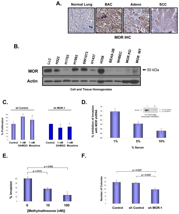
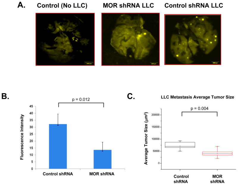
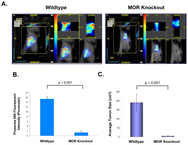
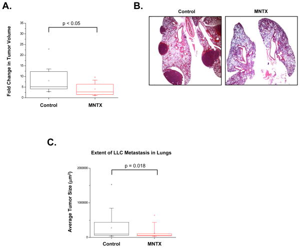
Results: We provide several lines of evidence that the MOR may be a potential target for lung cancer, a disease with high mortality and few treatment options. We first observed that there is ∼5- to 10-fold increase in MOR expression in lung samples from patients with NSCLC and in several human NSCLC cell lines. The MOR agonists morphine and [D-Ala(2), N-MePhe(4), Gly-ol]-enkephalin (DAMGO) increased in vitro LLC cell growth. Treatment with MNTX or silencing MOR expression inhibited LLC invasion and anchorage-independent growth by 50%-80%. Injection of MOR silenced LLC lead to a ∼65% reduction in mouse lung metastasis. In addition, MOR knockout mice do not develop significant tumors when injected with LLC in comparison with wild-type controls. Finally, continuous infusion of the peripheral opioid antagonist MNTX attenuates primary LLC tumor growth and reduces lung metastasis.

Conclusions: Taken together, our data suggest a possible direct effect of opiates on lung cancer progression, and provide a plausible explanation for the epidemiologic findings. Our observations further suggest a possible therapeutic role for opioid antagonists.


Conflict of interest statement

Competing Financial Interest

Dr. Moss serves as a paid consultant to Progenics Pharmaceuticals, Inc., has a financial interest in methylnaltrexone as a patent holder through the University of Chicago, and receives stock options from Progenics.

Figures

Figure 1
Figure 1. The mu opioid receptor (MOR) is overexpressed in human non-small cell lung cancer (NSCLC) and regulates Lewis lung carcinoma (LLC) oncogenic properties in vitro.
Panel A: Representative immunohistochemical (IHC) analysis of normal lung and lung tumor samples from patients indicate increased expression of MOR in bronchioloalveolar carcinoma (BAC), adenocarcinoma (Adeno) and, to a lesser extent, squamous cell carcinoma (SCC). MOR-specific brown staining intensity from 10 patient samples per group were analyzed with a statistically significant difference (p < 0.05) between normal and BAC, Adeno or SCC. Panel B: MOR immunoblot analysis of cell lysates from human NSCLC adneocarcinoma cell lines (H522, H1703, H1993, H1437), human NSCLC bronchioloalveolar carcinoma cell lines (SW1573, H358), non-cancerous BEAS-2B, normal human bronchial epithelial cells (NHBEC) and tissue homogenates from MOR knockout mouse brain and C57BL/6 mouse brain. There is increased MOR expression in several human NSCLC cell lines. Panel C: Stable control shRNA or MOR-1 shRNA-transfected LLC cells were analyzed for mu opioid agonist-mediated proliferation using a MTS proliferation assay. There is a statistically significant difference (p < 0.05, indicated by an asterisks) between control and DAMGO (1 nM, 24 hous) or morphine (1 nM, 24 hours)-treated LLC cells with n = 3 per condition. Stable silencing (shRNA) of the mu opioid receptor blocks mu opioid agonist-induced LLC proliferation. See the Methods section for experimental details. Panel D: Lewis lung carcinoma (LLC) cells were either transfected with control shRNA or MOR-1 shRNA, selected with puromycin and MOR protein expression analyzed (Panel C-inset). Cells were then analyzed for in vitro invasion with media containing 1, 5 or 10% serum, n = 3 per condition. There is a significant reduction in invasion (p < 0.05) between control shRNA and MOR-1 shRNA transfected cells for all conditions tested. Panel E: The peripheral MOR antagonist, methylnaltrexone (MNTX), inhibits LLC cell invasion. LLC cells were treated with 0, 10 or 100 nM MNTX and invasion assays performed. There is a statistically significant difference (p < 0.05) between groups with n = 3 per condition. Panel F: Stable control shRNA or MOR-1 shRNA-transfected LLC cells were analyzed for anchorage-independent growth using a soft agar colony formation assay. There is a statistically significant difference (p < 0.05) between groups with n = 3 per condition. See the Methods section for experimental details.
Figure 2
Figure 2. Silencing (shRNA) MOR expression in LLC inhibits tumor growth and metastasis
Panel A: Stable control shRNA or MOR-1 shRNA-transfected LLC cells were generated and either no cells (control), or LLC cells were injected (1×10⁵) intravenously. Mice were imaged at 4 weeks post injection with OV-100. Panel B: Graphical representation of quantitation of fluorescent intensity from experiments described in Panel A (p = 0.012, n = 4 mice per condition). Panel C: Graphical representation of quantitation of H & E stained sections of experiments similar to those described in Panel A. MOR silenced LLC cells exhibited a significant reduction in tumor metastasis volume (p = 0.004, n = 6 mice per condition). See the Methods section for experimental details.
Figure 3
Figure 3. Inhibition of LLC tumor formation in the mu opioid receptor (MOR) knockout mouse
C57BL/6 wildtype (WT) and mu opioid receptor knockout mice (MOR−/−) were injected with dual color labeled LLC cells (1×10⁶) subcutaneously in the right flank. Panel A: Visen FMT imaging of tumor volume at 12 days post visible tumor formation in wildtype C57BL/6 mouse (Left Panel) or MOR knockout mouse (Right Panel) after injecting Prosense680 (i.v, 2nM, 24 hours prior imaging) near infrared probe for quantification of tumor volume and lung metastasis. Panel B: Quantitation of Prosense680 fluorescence intensity in wildtype and MOR knockout mice (p < 0.001, n = 5 animals per condition). Panel C: Graphical analysis of LLC tumor volume in wildtype and MOR knockout mice (p < 0.001, n = 5 animals per condition). See the Methods section for experimental details.
Figure 4
Figure 4. Continuous infusion of MNTX inhibits LLC tumor growth and metastasis
Panel A: Graph indicating continuous infusion of MNTX (10 mg/kg/day, Alzet mini pumps) for two weeks after visible tumor formation in mice (LLC cells (1×10⁶) injected subcutaneously in the right flank) attenuates further tumor growth (n = 5 mice per condition, p < 0.05). Panel B: The same mice as described in Panel A were evaluated for lung metastasis from the primary flank tumor using H & E staining of excised formalin-fixed lung sections. Panel C: Graphical representation of the extent of lung metastasis with or without MNTX infusion, n = 5 mice per condition, p = 0.018. See the Methods section for experimental details.

Similar articles

Cited by

References

    1. Gottschalk A, Sharma S, Ford J, Durieux ME, Tiouririne M. Review article: the role of the perioperative period in recurrence after cancer surgery. Anesth Analg. 2010;110:1636–1643. - PubMed
    1. Biki B, Mascha E, Moriarty DC, Fitzpatrick JM, Sessler DI, Buggy DJ. Anesthetic technique for radical prostatectomy surgery affects cancer recurrence: a retrospective analysis. Anesthesiology. 2008;109:180–187. - PubMed
    1. Exadaktylos AK, Buggy DJ, Moriarty DC, Mascha E, Sessler DI. Can anesthetic technique for primary breast cancer surgery affect recurrence or metastasis? Anesthesiology. 2006;105:660–664. - PMC - PubMed
    1. Moss J, Israel RJ. Effects of anesthetics on cancer recurrence. J Clin Oncol. 2009;27:e89. author reply e90. - PubMed
    1. Singleton PA, Lingen MW, Fekete MJ, Garcia JG, Moss J. Methylnaltrexone inhibits opiate and VEGF-induced angiogenesis: Role of receptor transactivation. Microvasc Res. 2006;72:3–11. - PubMed

Publication types

Substances