. 2011 Jan;121(1):422-30.
doi: 10.1172/JCI44244. Epub 2010 Dec 13.

Cardiac neural crest orchestrates remodeling and functional maturation of mouse semilunar valves

Affiliations

Cardiac neural crest orchestrates remodeling and functional maturation of mouse semilunar valves

Rajan Jain et al. J Clin Invest. 2011 Jan.

Abstract

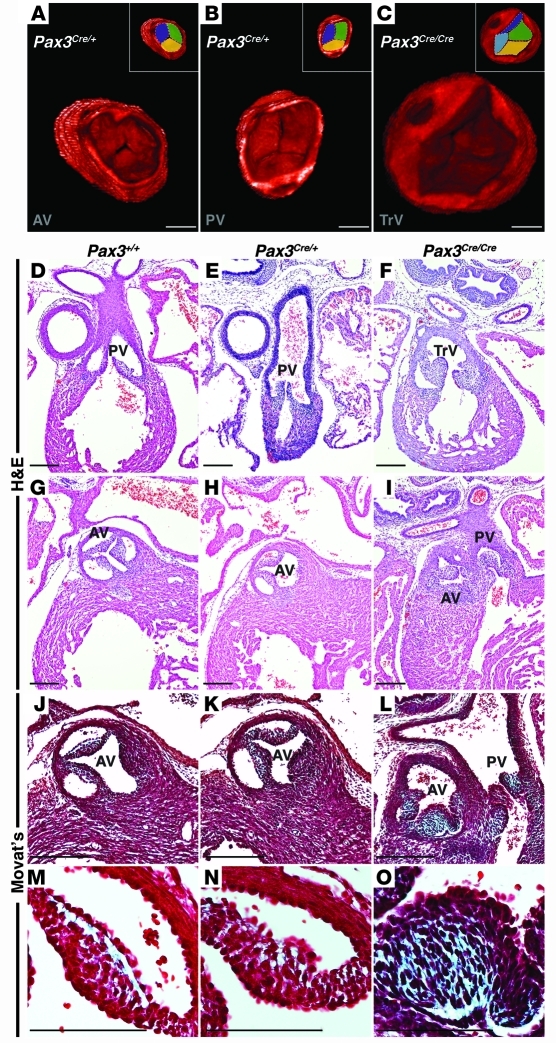
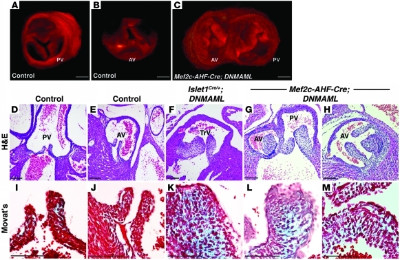
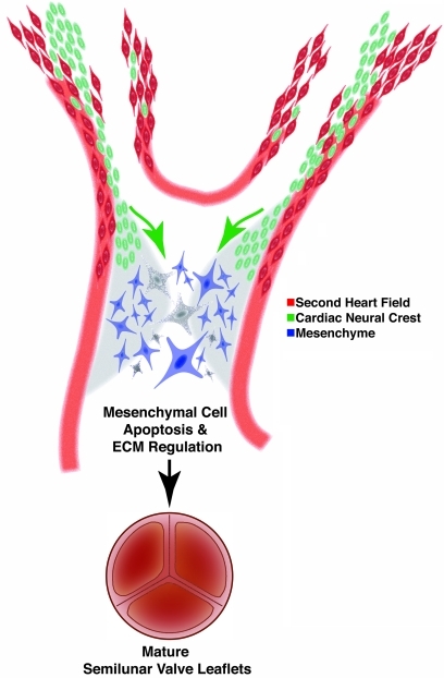
Congenital anomalies of the aortic valve are common and are associated with progressive valvular insufficiency and/or stenosis. In addition, aneurysm, coarctation, and dissection of the ascending aorta and aortic arch are often associated conditions that complicate patient management and increase morbidity and mortality. These associated aortopathies are commonly attributed to turbulent hemodynamic flow through the malformed valve leading to focal defects in the vessel wall. However, numerous surgical and pathological studies have identified widespread cystic medial necrosis and smooth muscle apoptosis throughout the aortic arch in affected patients. Here, we provide experimental evidence for an alternative model to explain the association of aortic vessel and valvular disease. Using mice with primary and secondary cardiac neural crest deficiencies, we have shown that neural crest contribution to the outflow endocardial cushions (the precursors of the semilunar valves) is required for late gestation valvular remodeling, mesenchymal apoptosis, and proper valve architecture. Neural crest was also shown to contribute to the smooth muscle layer of the wall of the ascending aorta and aortic arch. Hence, defects of cardiac neural crest can result in functionally abnormal semilunar valves and concomitant aortic arch artery abnormalities.


Figures

Figure 1
Figure 1. Loss of Pax3 results in abnormal semilunar valve leaflets.
3D reconstruction of OPT images of E16.5 Pax3Cre/+ control embryos demonstrates a trileaflet aortic (A) and pulmonic (B) valve, each with 3 commissures, while Pax3Cre/Cre mutant embryos demonstrate abnormal semilunar valve leaflets. The mutant depicted in C displays a truncal valve with 4 leaflets. Insets in AC represent identical images with each leaflet pseudocolored. Cross-sectional H&E images through the pulmonic valve leaflets of E16.5 Pax3+/+ and Pax3Cre/+ embryos (D and E) and a Pax3Cre/Cre littermate (F) show thickened leaflets in the mutant. Cross-sectional H&E images through the aortic valve leaflets of E16.5 Pax3+/+ and Pax3Cre/+ embryos (G and H) and a Pax3Cre/Cre littermate (I) show thickened, unequally sized leaflets in the mutant. Examples of a truncal valve (F) and double-outlet right ventricle (I) are shown. Modified Movat’s Pentachrome staining reveals extracellular matrix deposition (blue) in E16.5 control semilunar valve leaflets (J and K) and Pax3Cre/Cre embryos (L), which show an increase in extracellular matrix compared with control leaflets. Higher magnification images of JL are shown as MO. AV, aortic valve; PV, pulmonic valve; TrV, truncal valve. Brightness and contrast of OPT images were adjusted using OsiriX software. Scale bars: 100 μm.
Figure 2
Figure 2. Loss of Pax3 results in aortic regurgitation.
Pulse-wave Doppler analysis from the aorta of E16.5 Pax3+/+ and Pax3Cre/+ control embryos demonstrate no significant aortic regurgitation (A and B). Pax3Cre/Cre littermates demonstrate at least moderate-severe aortic insufficiency (arrowhead in C and D). Arrowheads point to regurgitant flow in mutant embryos.
Figure 3
Figure 3. Abnormal neural crest migration in neural crest and second heart field mutants.
In situ hybridization for neural crest marker Sema3C of E11.5 coronal sections through the outflow tract cushions of control (WT) (A), Pax3Cre/Cre (B), Islet1Cre/+;DNMAML (C), and Mef2c-AHF-Cre;DNMAML (D) embryos. In situ hybridization for neural crest marker PlexinA2 of E11.5 coronal sections of control (WT) (E), Pax3Cre/Cre (F), Islet1Cre/+;DNMAML (G), and Mef2c-AHF-Cre;DNMAML (H) embryos. AP2α immunohistochemistry of coronal sections of the outflow tract cushions of E10.5 control (WT) (I) and Mef2c-AHF-Cre;DNMAML (J) embryos. Dotted lines in A, E, and I enclose regions occupied by neural crest cells expressing Sema3C, PlexinA2, and AP2α, respectively. GFP and Islet1 immunohistochemistry of cross sections through the outflow tract and pharyngeal mesenchyme of E9.5 Pax3Cre/+;Z/EG embryos (K and L). GFP-positive (neural crest derivatives) and Islet1-positive (second heart field) cells that are in close apposition to one another are highlighted with green (GFP) or red (Islet1) arrowheads. Scale bars: 100 μm.
Figure 4
Figure 4. Notch inhibition in the second heart field results in dysmorphic semilunar valve leaflets.
3D reconstructions of OPT-generated images of E17.5 control (WT) pulmonic valve (A), aortic valve (B), and Mef2c-AHF-Cre;DNMAML semilunar valve leaflets (C). Note the thickened aortic valve leaflets and bicuspid pulmonic valve in the mutant (C), which demonstrates a double-outlet right ventricle. H&E staining of cross sections of E17.5 control (WT) pulmonic (D) and aortic valves (E) demonstrates delicate, thin leaflets. However, Islet1Cre/+;DNMAML (F) and Mef2c-AHF-Cre;DNMAML semilunar valve leaflets (G and H) are thickened and of unequal sizes. Modified Movat’s Pentachrome staining of cross sections of E17.5 control (WT) pulmonic (I), aortic (J), Islet1Cre/+;DNMAML (K), and Mef2c-AHF-Cre;DNMAML valve leaflets (L and M). Mutant leaflets demonstrate an increase in extracellular matrix deposition. F and K show embryos with a persistent truncus arteriosus, and G, H, L, and M show embryos with a double-outlet right ventricle. Brightness and contrast of OPT images were adjusted using OsiriX software. Scale bars: 100 μm.
Figure 5
Figure 5. Inhibition of Notch signaling in the second heart field results in aortic regurgitation.
Pulse-wave Doppler analysis from the aorta of E17.5 control (A), Islet1Cre/+;DNMAML (B), control (C), and Mef2c-AHF-Cre;DNMAML (D) embryos. Control embryos are WT littermates of the respective mutants. Arrowheads point to regurgitant flow in mutant embryos.
Figure 6
Figure 6. Loss of Pax3 or impaired second heart field Notch signaling results in diminished mesenchymal apoptosis in late gestation semilunar valve leaflets.
Quantification of TUNEL+ cells (as a percentage of all DAPI+ mesenchymal cells) in E16.5 semilunar valve leaflets of Pax3 mutants (A). At least 6 sections were analyzed from each of 3 embryos for each genotype. Total number of cells counted are as follows: Pax3+/+, 6495 cells; Pax3Cre/+, 7314 cells; and Pax3Cre/Cre, 8749 cells. E17.5 semilunar valve leaflets of control (WT) and Mef2c-AHF-Cre;DNMAML embryos (B). At least 8 sections were analyzed from each of 3 embryos for each genotype. Total number of cells counted are as follows: control, 15445 cells; and Mef2c-AHF-Cre;DNMAML, 40707 cells. Each point represents the percentage of TUNEL+ cells in an individual embryo.
Figure 7
Figure 7. Model depicting the role of neural crest in semilunar valve development.
Neural crest cells (green) are in close apposition to second heart field precursors (red) in the ventral pharynx during migration to the outflow endocardial cushions (light gray), where they provide instructive signals to orchestrate apoptosis (depicted as dark gray cells) and changes in extracellular matrix production during valve remodeling.

References

    1. Nakajima Y, Yamagishi T, Hokari S, Nakamura H. Mechanisms involved in valvuloseptal endocardial cushion formation in early cardiogenesis: roles of transforming growth factor (TGF)-beta and bone morphogenetic protein (BMP). Anat Rec. 2000;258(2):119–127. doi: 10.1002/(SICI)1097-0185(20000201)258:2<119::AID-AR1>3.0.CO;2-U. - DOI - PubMed
    1. Butcher JT, Markwald RR. Valvulogenesis: the moving target. Philos Trans R Soc Lond B Biol Sci. 2007;362(1484):1489–1503. - PMC - PubMed
    1. Combs MD, Yutzey KE. Heart valve development: regulatory networks in development and disease. Circ Res. 2009;105(5):408–421. doi: 10.1161/CIRCRESAHA.109.201566. - DOI - PMC - PubMed
    1. Markwald RR, Norris RA, Moreno-Rodriguez R, Levine RA. Developmental basis of adult cardiovascular diseases: valvular heart diseases. Ann N Y Acad Sci. 2010;1188:177–183. doi: 10.1111/j.1749-6632.2009.05098.x. - DOI - PMC - PubMed
    1. Fedak PW, Verma S, David TE, Leask RL, Weisel RD, Butany J. Clinical and pathophysiological implications of a bicuspid aortic valve. Circulation. 2002;106(8):900–904. doi: 10.1161/01.CIR.0000027905.26586.E8. - DOI - PubMed

Publication types

MeSH terms