. 2011 Jan;121(1):265-76.
doi: 10.1172/JCI42596. Epub 2010 Dec 13.

Bcl3 prevents acute inflammatory lung injury in mice by restraining emergency granulopoiesis

Affiliations

Bcl3 prevents acute inflammatory lung injury in mice by restraining emergency granulopoiesis

Daniel Kreisel et al. J Clin Invest. 2011 Jan.

Abstract

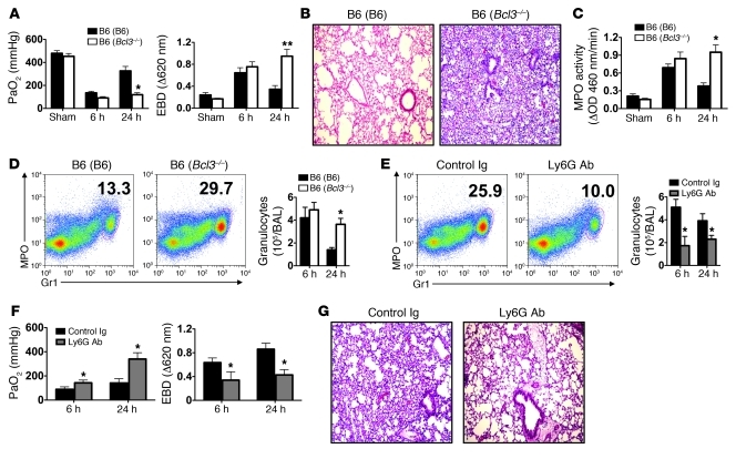
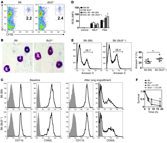
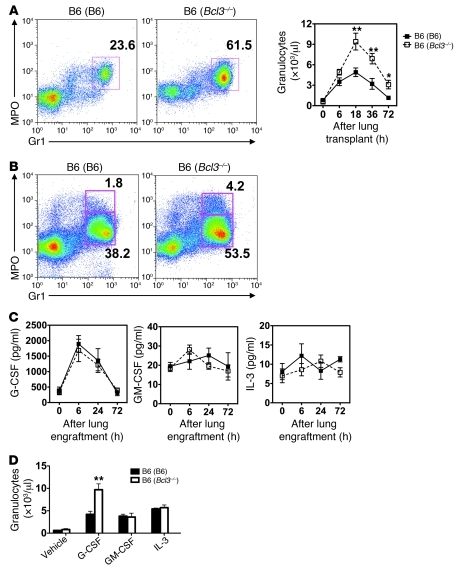
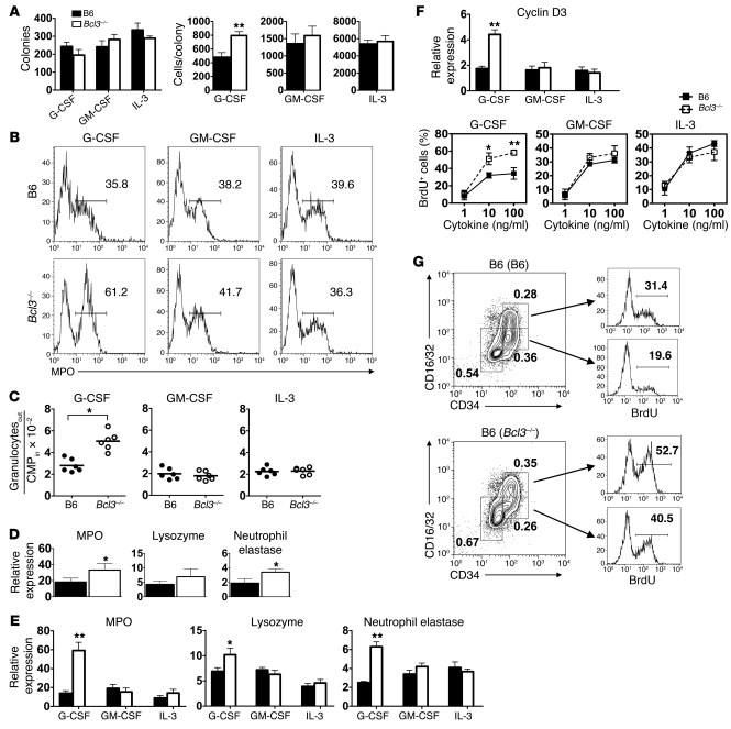
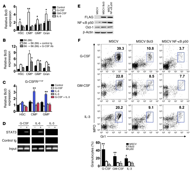
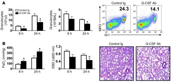
Granulocytes are pivotal regulators of tissue injury. However, the transcriptional mechanisms that regulate granulopoiesis under inflammatory conditions are poorly understood. Here we show that the transcriptional coregulator B cell leukemia/lymphoma 3 (Bcl3) limits granulopoiesis under emergency (i.e., inflammatory) conditions, but not homeostatic conditions. Treatment of mouse myeloid progenitors with G-CSF--serum concentrations of which rise under inflammatory conditions--rapidly increased Bcl3 transcript accumulation in a STAT3-dependent manner. Bcl3-deficient myeloid progenitors demonstrated an enhanced capacity to proliferate and differentiate into granulocytes following G-CSF stimulation, whereas the accumulation of Bcl3 protein attenuated granulopoiesis in an NF-κB p50-dependent manner. In a clinically relevant model of transplant-mediated lung ischemia reperfusion injury, expression of Bcl3 in recipients inhibited emergency granulopoiesis and limited acute graft damage. These data demonstrate a critical role for Bcl3 in regulating emergency granulopoiesis and suggest that targeting the differentiation of myeloid progenitors may be a therapeutic strategy for preventing inflammatory lung injury.


Figures

Figure 1
Figure 1. Granulocytes promote lung graft injury in Bcl3-deficient recipients.
(A) Assessment of PaO2 (left) and exclusion of EBD (right) 6 and 24 hours following B6 → B6 (B6) or B6 → B6 (Bcl3–/–) lung engraftment. (B and C) Representative graft histology (original magnification, ×100) (n = 11) (B) and intragraft MPO activity (C) 24 hours following B6 → B6 (B6) or B6 → B6 (Bcl3–/–) lung engraftment. (D) Left: Representative FACS analysis (n = 5). Numbers denote percent abundance of granulocytes in graft tissue. Right: Granulocyte counts in BAL at 6 and 24 hours following B6 → B6 (B6) or B6 → B6 (Bcl3–/–) lung engraftment. (E) Left: B6 → B6 (Bcl3–/–) lung recipients were partially depleted for granulocytes with Ly6G-specific antibodies or treated with control Ig 4 hours prior to transplantation and assessed for percent abundance of granulocytes in graft tissue 24 hours after engraftment. Right: Granulocyte counts at 6 and 24 hours following engraftment in BAL. (F) Assessment of PaO2 (left) and EBD exclusion (right) at 6 and 24 hours following engraftment. (G) Representative lung graft histology (original magnification, ×100) of B6 → B6 (Bcl3–/–) recipients 24 hours after engraftment following treatment with either control Ig or Ly6G-specific antibodies (n = 4). Data are mean ± SD and, unless otherwise indicated, represent at least 3 independent experiments. *P < 0.05; **P < 0.01.
Figure 2
Figure 2. Functional and phenotypic assessment of B6 and Bcl3-deficient granulocytes.
(A) Assessment of granulocyte recruitment into lung grafts. CFSE-labeled B6 or Bcl3–/– granulocytes (5 × 10⁶) were assessed by FACS analysis in B6 → B6 lung grafts 3 hours following adoptive transfer. Numbers denote percent abundance of CFSE-labeled granulocytes within grafts. Results are representative of 3 independent experiments. (B) Nuclear morphology (original magnification, ×400) of B6 or Bcl3–/– granulocytes following Wright Giemsa stain. (C) Evaluation of granulocyte activation measured by CD11b and CD62L expression on granulocytes before (left) or 18 hours after (right) B6 → B6 (B6) or B6 → B6 (Bcl3–/–) lung engraftment. Results are representative of 5 independent experiments. (D) Measurement of f-MLP– or PMA-mediated ROS generation from bone marrow–derived or BAL-derived B6 or Bcl3–/– granulocytes 24 hours after engraftment. Data are representative of 3 independent experiments. (E) Survival in vivo of BAL granulocytes from B6 → B6 (Bcl3–/–) or B6 → B6 (B6) lung recipients 24 hours following transplantation depicted as a representative FACS plot (n = 6) (left) or as a scatter plot (right) showing percent abundance of Annexin V+ granulocytes. Data are representative of 7 independent experiments. (F) Survival ex vivo of B6 or Bcl3–/– granulocytes in the absence or presence of G-CSF (10 ng/ml) shown normalized to the number of Annexin V cells at the initiation of culture. Data are representative of 2 independent experiments. Data represent mean ± SD. *P < 0.05.
Figure 3
Figure 3. Bcl3 limits G-CSF–mediated emergency granulopoiesis.
(A) Left: Representative FACS analysis (n = 3). Numbers denote percent abundance of peripheral blood granulocytes at 24 hours following B6 → B6 (Bcl3–/–) or B6 → B6 (B6) lung engraftment. Right: Peripheral blood granulocyte numbers for indicated time points up to 72 hours after B6 → B6 (Bcl3–/–) or B6 → B6 (B6) lung transplantation (n = 4). (B) Representative FACS analysis (n = 3) of MPO+Gr1+ populations in the bone marrow of B6 (B6) or B6 (Bcl3–/–) mice 18 hours following the implantation of B6 lungs. Numbers indicate percent abundance of the indicated populations within the bone marrow. (C) Serum concentrations of indicated cytokines following B6 → B6 (Bcl3–/–) or B6 → B6 (B6) lung engraftment (n = 4). (D) Peripheral blood granulocyte numbers 24 hours following intravenous administration of 5 μg of G-CSF, GM-CSF, or IL-3 to either B6 (B6) or B6 (Bcl3–/–) mice (n = 5). Data represent mean ± SD. *P < 0.05; **P < 0.01.
Figure 4
Figure 4. Proliferation and differentiation of Bcl3-deficient myeloid progenitors.
(A) Left: Methylcellulose colony count from B6 or Bcl3–/– bone marrow cells cultured with indicated cytokines. Right: Number of cells per colony. Data are representative of 2 independent experiments. (B) Representative FACS analysis (n = 4) of MPO expression in liquid CMP cultures following 18 hours of stimulation with indicated cytokines. Numbers within histograms indicate percent abundance of MPO+ cells. (C) Granulocyte output from CMP liquid cultures following 72 hours of stimulation with indicated cytokines. (D) Indicated transcript accumulation in peripheral blood granulocytes isolated from B6 (B6) or B6 (Bcl3–/–) mice 24 hours following a 5-μg injection of G-CSF. (E) Indicated transcript accumulation in CMPs isolated from B6 (B6) or B6 (Bcl3–/–) mice 24 hours following injection of 5 μg of indicated cytokines. (F) Bottom: CMP BrdU incorporation in response to indicated concentrations of G-CSF, GM-CSF, or IL-3 following 18 hours of liquid culture. Data are representative of at least 3 independent experiments. Top: Cyclin D3 transcript accumulation in CMPs isolated from B6 (B6) or B6 (Bcl3–/–) mice 24 hours following injection of 5 μg of indicated cytokines. (G) Representative (n = 3) BrdU incorporation of bone marrow LinSca-1c-Kit+ cells (GMPs; CD34+CD16/32+) and (CMPs; CD34+CD16/32) in B6 →B6 (B6) or B6 (Bcl3–/–) lung recipients 24 hours following transplantation. Dot plot numbers indicate percent abundance. Data represent mean ± SD. *P < 0.05; **P < 0.01.
Figure 5
Figure 5. The dynamics and effects of Bcl3 expression in myeloid progenitors.
(A) Representative (n = 4) Bcl3 transcript expression in B6 myeloid progenitors or granulocytes (Gran) before (control) and after 18 hours of stimulation with 10 ng/ml of indicated cytokines in liquid culture. (B) Representative (n = 4) Bcl3 transcript level expression in myeloid progenitors and granulocytes purified from resting B6 mice, B6 → B6 (B6) treated with control Ig or G-CSF–specific antibodies 18 hours following transplantation. (C) Representative (n = 2) Bcl3 transcript accumulation in G-CSFRΔ715F myeloid cell progenitors. (D) Representative (n = 2) analysis of STAT3 association with Bcl3 promoter. Lin B6 bone marrow cells were stimulated with indicated cytokines. Chromatin immunoprecipitation was then conducted with STAT3-specific or control antibodies, and amplification was performed with primers specific for an enhancer region of Bcl3. (E) Assessment of Bcl3 ectopic expression on NF-κB p50 protein accumulation. Lin B6 bone marrow cells were transfected with MSCV, MSCV-Bcl3 (encoding N-FLAG Bcl3), or MSCV NF-κB p50 (encoding N-FLAG NF-κB p50). Nuclear protein was extracted, immunoblotted, and probed with FLAG–, NF-κB p50–, Oct-1–, and β-actin–specific antibodies. Results are representative of 3 independent experiments. (F) Top: Representative FACS analysis (n = 5). Numbers denote percent abundance of granulocytes in Lin bone marrow cell cultures following 3 days of stimulation with indicated cytokines. Bottom: Mean percent abundance of granulocytes calculated from 5 independently conducted cultures derived from data in top panel. Data represent mean ± SD. *P < 0.05; **P < 0.01.
Figure 6
Figure 6. G-CSF blockade prevents graft injury in B6 (Bcl3–/–) lung recipients.
(A) Granulocyte numbers in the peripheral blood (left) and BAL (middle) at 6 and 24 hours post-engraftment and a representative FACS analysis (24 hours; right) in which numbers indicate granulocyte percent abundance in graft tissue in B6 → B6 (Bcl3–/–) lung recipients treated with control Ig or G-CSF–specific antibodies (n = 10). (B) PaO2 (left) and EBD exclusion (middle) 6 and 24 hours post-engraftment and representative graft histology (right; original magnification, ×100) in B6 → B6 (Bcl3–/–) lung graft recipients 24 hours after engraftment treated with control Ig or G-CSF–specific antibodies (n = 10). Data represent mean ± SD. *P < 0.05.

References

    1. Weiss SJ. Tissue destruction by neutrophils. N Engl J Med. 1989;320(6):365–376. doi: 10.1056/NEJM198902093200606. - DOI - PubMed
    1. Martin P, et al. Wound healing in the PU.1 null mouse--tissue repair is not dependent on inflammatory cells. Curr Biol. 2003;13(13):1122–1128. doi: 10.1016/S0960-9822(03)00396-8. - DOI - PubMed
    1. Watari K, et al. Serum granulocyte colony-stimulating factor levels in healthy volunteers and patients with various disorders as estimated by enzyme immunoassay. Blood. 1989;73(1):117–122. - PubMed
    1. Cheers C, Haigh AM, Kelso A, Metcalf D, Stanley ER, Young AM. Production of colony-stimulating factors (CSFs) during infection: separate determinations of macrophage-, granulocyte-, granulocyte-macrophage-, and multi-CSFs. Infect Immun. 1988;56(1):247–251. - PMC - PubMed
    1. Metcalf D, Begley CG, Johnson GR, Nicola NA, Lopez AF, Williamson DJ. Effects of purified bacterially synthesized murine multi-CSF (IL-3) on hematopoiesis in normal adult mice. Blood. 1986;68(1):46–57. - PubMed

Publication types

MeSH terms