

Case Reports
doi: 10.4103/0301-4738.73721.
Clinical features and management of posttraumatic subperiosteal hematoma of the orbit
Affiliations
- PMID: 21157076
- PMCID: PMC3032247
- DOI: 10.4103/0301-4738.73721
Item in Clipboard
Case Reports
Clinical features and management of posttraumatic subperiosteal hematoma of the orbit
Indian J Ophthalmol.
2011 Jan-Feb.
Abstract
Traumatic subperiosteal hematoma (SpH) usually presents late, after the initial trauma. It is generally seen in young males. Computed tomography is the best mode of imaging and helps to rule out orbital fracture or associated subdural hematoma. We present the clinical features and management of four patients seen at the orbit clinic with SpH. Management is based on time of presentation, visual acuity and any communicating bleed. The prognosis of traumatic SpH is excellent if treated with an individualized patient approach.
Figures
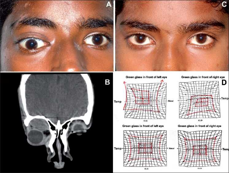
(A) Clinical photograph of the patient showing proptosis and downward displacement of the globe. (B) Computerized tomography scan photograph (coronal) showing a hypodense lesion measuring 3 × 2 × 1.2 cm superior to the optic nerve and superior rectus muscle. (C) Clinical photograph of the same patient 1 month after treatment of steroids showing complete resolution of proptosis. (D) Hess charting upper image showing restriction of movements in the right eye at the time of presentation and lower image showing normal movements after treatment

(A) Clinical photograph of the patient showing proptosis and downward displacement of the globe. (B) Hess charting showing restriction of movements in the right eye. (C and D) Computerized tomography scan photograph (coronal and sagittal) showing a hypodense lesion in the superior quadrant of the right orbit measuring 3.2 × 2.7 × 1.4 cm, pushing the globe down and out. (E) Photograph of the same patient 1 week after aspiration of the hematoma. (F) Hess charting showing normal movements after treatment

(A and B) Clinical photograph of the patient showing the nonaxial proptosis and exposure keratopathy in the left eye with a lacerated wound over the left supraorbital ridge. (C, D and E) Computerized tomography scan (axial and coronal) showing superomedial orbital subperiosteal hematoma measuring 3.44 × 2.73 × 2.24 cm, in continuity with the subfrontal extradural hematoma measuring 5.75 × 4.53 × 3.30 cm through the undisplaced orbital roof fracture in the left side and displacing the globe downwards, laterally and forward. (F) Postoperative photograph 1 week after frontal craniotomy and superior orbitotomy showing complete resolution of proptosis
References
-
- Choi HY, Han YS, Lee JS, Oum BS. Subperiosteal hematoma after surgical treatment for subarachnoid hemorrhage. Ophthal Plast Reconstr Surg. 2004;20:87–8. - PubMed
-
- Woo KI, Kim YD. Subperiosteal hematoma of the orbit associated with sinusitis. Korean J Ophthalmol. 1997;11:118–22. - PubMed
-
- Yazici B, Taner P. Delayed orbital subperiosteal hemorrhage after blunt trauma. Ophthal Plast Reconstr Surg. 2007;23:315–6. - PubMed
-
- Seigel RS, Williams AG, Hutchison JW, Wolter JR, Carlow TJ, Rogers DE. Subperiosteal hematomas of the orbit: angiographic and computed tomographic diagnosis. R1982;143:711–4. - PubMed
Publication types
MeSH terms
LinkOut - more resources
Full Text Sources
