

Combination photodynamic therapy and bevacizumab for choroidal neovascularization associated with toxoplasmosis
- PMID: 21157079
- PMCID: PMC3032250
- DOI: 10.4103/0301-4738.73728
Combination photodynamic therapy and bevacizumab for choroidal neovascularization associated with toxoplasmosis
Abstract
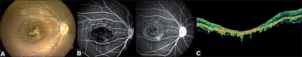
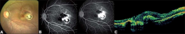
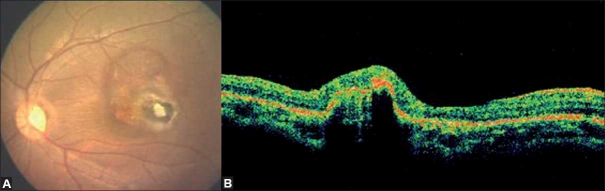
A 14-year-old girl presenting with visual loss in both eyes was diagnosed to have healed toxoplasma retinochoroiditis in the right eye with active choroidal neovascularization (CNV) secondary to toxoplasmosis in the left. She underwent combination photodynamic therapy (PDT) and intravitreal bevacizumab as primary treatment. PDT was performed as per the 'Treatment of Age-related Macular Degeneration by Photodynamic therapy' study protocol and was followed by intravitreal bevacizumab after 2 days. CNV regressed at 8 weeks of follow-up and remained stable at 8 months of follow-up. The initial visual acuity improved from 20/120 to 20/30. Combination therapy with PDT and intravitreal bevacizumab appears to be effective in the treatment of CNV secondary to toxoplasma retinochoroiditis.
Figures
Similar articles
-
Intravitreal bevacizumab for choroidal neovascularization in toxoplasmosis.Acta Ophthalmol. 2009 Sep;87(6):688-90. doi: 10.1111/j.1755-3768.2008.01319.x. Epub 2008 Jul 8. Acta Ophthalmol. 2009. PMID: 18631326 No abstract available.
-
Bevacizumab vs photodynamic therapy for choroidal neovascularization in multifocal choroiditis.Arch Ophthalmol. 2010 Sep;128(9):1100-3. doi: 10.1001/archophthalmol.2010.205. Arch Ophthalmol. 2010. PMID: 20837791 Clinical Trial.
-
Pattern electroretinographic results after photodynamic therapy alone and photodynamic therapy in combination with intravitreal bevacizumab for choroidal neovascularization in age-related macular degeneration.Doc Ophthalmol. 2009 Aug;119(1):37-42. doi: 10.1007/s10633-009-9167-8. Epub 2009 Feb 19. Doc Ophthalmol. 2009. PMID: 19225818
-
Intravitreal injection of bevacizumab combined with verteporfin photodynamic therapy for choroidal neovascularization in age-related macular degeneration.Retina. 2008 May;28(5):675-81. doi: 10.1097/IAE.0b013e31816b316e. Retina. 2008. PMID: 18463509
-
Intravitreal bevacizumab for treatment of choroidal neovascularization secondary to toxoplasmic retinochoroiditis: a case series.Semin Ophthalmol. 2015 May;30(3):181-7. doi: 10.3109/08820538.2013.838278. Epub 2013 Oct 31. Semin Ophthalmol. 2015. PMID: 24175641
Cited by
-
Choroidal neo-vascular membrane in a paediatric optic disc pit: A case report.Am J Ophthalmol Case Rep. 2022 Nov 12;28:101751. doi: 10.1016/j.ajoc.2022.101751. eCollection 2022 Dec. Am J Ophthalmol Case Rep. 2022. PMID: 36439653 Free PMC article.
-
An update on inflammatory choroidal neovascularization: epidemiology, multimodal imaging, and management.J Ophthalmic Inflamm Infect. 2018 Sep 12;8(1):13. doi: 10.1186/s12348-018-0155-6. J Ophthalmic Inflamm Infect. 2018. PMID: 30209691 Free PMC article. Review.
-
Toxoplasma Retinochoroiditis with Chorioretinal Neovascularization in a Young Patient.Case Rep Ophthalmol. 2021 Apr 16;12(1):259-263. doi: 10.1159/000512286. eCollection 2021 Jan-Apr. Case Rep Ophthalmol. 2021. PMID: 34054467 Free PMC article.
-
Clinical outcomes of intravitreal treatment for ocular toxoplasmosis: systematic review and meta-analysis.Rev Soc Bras Med Trop. 2023 May 22;56:e05522022. doi: 10.1590/0037-8682-0552-2022. eCollection 2023. Rev Soc Bras Med Trop. 2023. PMID: 37222350 Free PMC article.
-
Inflammatory choroidal neovascularization in Indian eyes: Etiology, clinical features, and outcomes to anti-vascular endothelial growth factor.Indian J Ophthalmol. 2017 Apr;65(4):295-300. doi: 10.4103/ijo.IJO_262_16. Indian J Ophthalmol. 2017. PMID: 28513493 Free PMC article.
References
-
- Sivaprasad S, Moore AT. Choroidal neovascularisation in children. Br J Ophthalmol. 2008;92:451–4. - PubMed
-
- Treatment of Age related macular degeneration with photodynamic therapy (TAP) study group: Photodynamic therapy of subfoveal CNV in ARMD with verteporfin; One year results of 2 Randomized clinical trials- TAP report. Arch Ophthalmol. 1999;117:1329–45. - PubMed
-
- Atmaca Ls, Simsek T, Batioglu F. Clinical features and prognosis in ocular toxoplasmosis. Jpn J Ophthalmol. 2004;48:386–91. - PubMed
-
- Augsburger JJ, Benson WE. Subretinal neovascularisation in chronic uveitis. Albrecht Von Graefes Arch Klin Exp Ophthalmol. 1980;215:43–51. - PubMed
-
- Goshorn EB, Hoover DL, Eller AW, Friberg TR, Jarrett WH, 2nd, Sorr EM. Subretinal revascularization in children and adolescents. J Paediatr Ophthalmol Strabismus. 1995;32:178–82. - PubMed
Publication types
MeSH terms
Substances
LinkOut - more resources
Full Text Sources
