Comparative Study
. 2011 Jan;70(1):69-82.
doi: 10.1097/NEN.0b013e318203ae74.

Defective glial maturation in vanishing white matter disease

Affiliations
Comparative Study

Defective glial maturation in vanishing white matter disease

Marianna Bugiani et al. J Neuropathol Exp Neurol. 2011 Jan.

Abstract

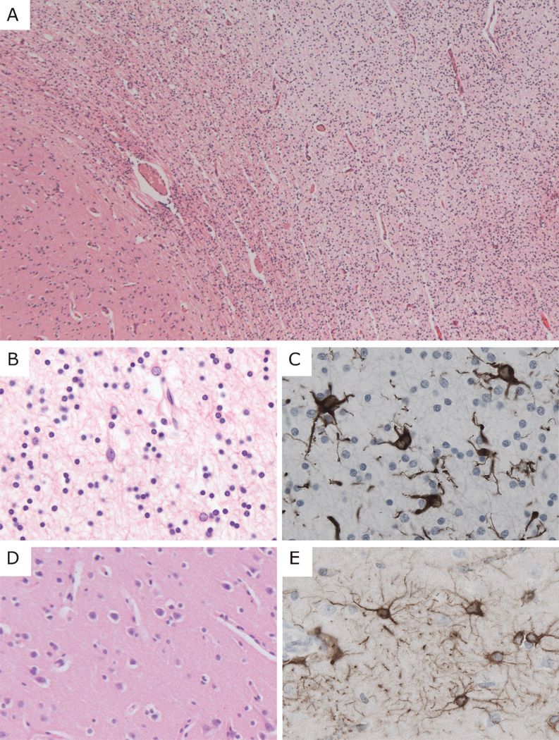
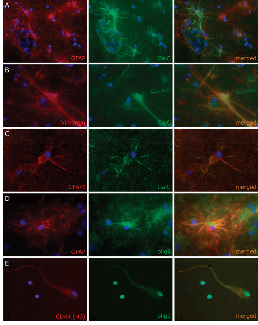
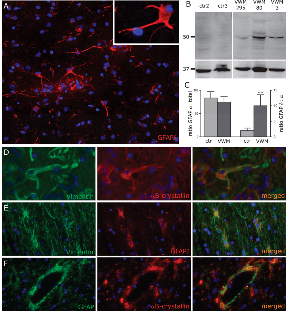
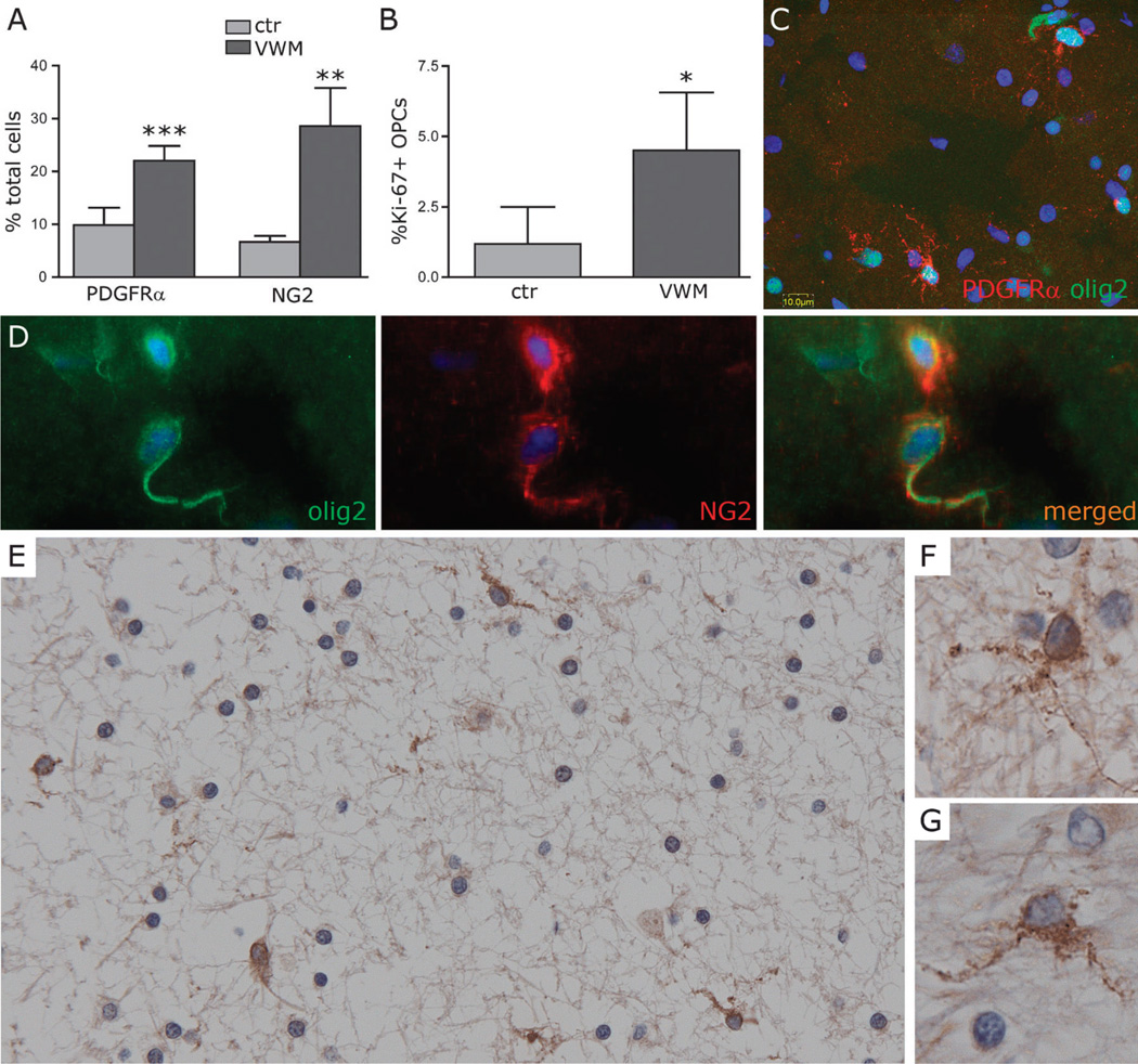
Vanishing white matter (VWM) disease is a genetic leukoencephalopathy linked to mutations in the eukaryotic translation initiation factor 2B. It is a disease of infants, children, and adults who experience a slowly progressive neurologic deterioration with episodes of rapid clinical worsening triggered by stress and eventually leading to death. Characteristic neuropathologic findings include cystic degeneration of the white matter with scarce reactive gliosis, dysmorphic astrocytes, and paucity of myelin despite an increase in oligodendrocytic density. To assess whether a defective maturation of macroglia may be responsible for the feeble gliosis and lack of myelin, weinvestigated the maturation status of astrocytes and oligodendrocytes in the brains of 8 VWM patients, 4 patients with other white matter disorders and 6 age-matched controls with a combination of immunocytochemistry, histochemistry, scratch-wound assays, Western blot, and quantitative polymerase chain reaction. We observed increased proliferation and a defect in the maturation of VWM astrocytes. They show an anomalous composition of their intermediate filament network with predominance of the δ-isoform of the glial fibrillary acidic protein and an increase in the heat shock protein αB-crystallin, supporting the possibility that a deficiency in astrocyte function may contribute to the loss of white matter in VWM. We also demonstrated a significant increase in numbers of premyelinating oligodendrocyte progenitors in VWM, which may explain the coexistence of oligodendrocytosis and myelin paucity in the patients' white matter.


Figures

Figure 1
Figure 1
General neuropathological features of vanishing white matter disease. (A) Low-power magnification hematoxylin and eosin staining of the white matter (right side of field) from the frontal lobe of patient VWM367 shows tissue rarefaction and increased cellular density. The U-fibers appear relatively preserved; the overlying cortex is normal. (B, C) At higher magnification, white matter astrocytes display abnormal morphology with coarse blunt processes. (D, E) The cortical architecture and morphology of cortical astrocytes are normal. (A, B, D) Hematoxylin and eosin; (C, E) immunohistochemistry for glial fibrillary acidic protein. Original magnifications: A, 25×; B–E, 200×.
Figure 2
Figure 2
Vanishing white matter disease (VWM) astrocytes proliferate actively but do not reach full maturity. (A) There are significantly higher numbers of Ki-67-positive astrocytes in the white matter of VWM patients than in controls (n = 5 patients, 3 controls; p < 0.0001; bars = SD). (B) Double stain for glial fibrillary acidic protein (GFAP) and Ki-67 shows cells with incomplete separation of the nucleus and abnormal morphology with blunt processes (patient VWM3). (C) Scratch-wound assays of control 5 (left panel) and patient VWM44 (right panel) show increased proliferation of primary cultured astrocytes in the scratch 48 hours after administration of a mechanical stress. Nuclei of the proliferating cells inside the scratch area are blue. Nuclei of the unstressed cells outside this area are red; their net number is not changed vs. controls. (D) The slope of proliferation in the scratch area; the individual net values are indicated as dots (n = 4 patients, 3 controls; p = 0.06; the lack of significance is due to the large variance of cell growth among the different VWM patients vs. controls). (E, F) GFAP-positive astrocytes from VWM patients express the intermediate filament nestin (patient VWM99) (E), and are S100β-negative (F) patient VWM3), suggesting that they are immature. (G) Astrocytes from controls show normal morphology with fine arborizations and both nuclear and cytoplasmic S100β-immunoreactivity (control 2; single S100β staining in inset). Nuclei are stained with DAPI (blue) in all panels. Original magnifications: (B, F) bars indicate 2 µm and 10 µm, respectively; original magnifications: E, G, 400×.
Figure 3
Figure 3
Vanishing white matter disease (VWM) astrocytes express oligodendrocytic lineage-specific proteins. (A, B) Double stain for glial fibrillary acidic protein (GFAP) (A, patient VWM3) or vimentin (B, patient VWM99) and galactocerebroside (GalC) show dysmorphic astrocytes with homogeneous cytoplasmic GalC immunoreactivity extending far into the processes. (C) In astrocytes from VWM patients, GalC colocalizes with other astrocytic lineage specific-markers, including GFAPδ (patient VWM44). (D) GFAP-positive cells with astrocyte morphology also express olig2 with a cytoplasmic immunoreactivity pattern (patient VWM80). E: Primary culture of sorted glial progenitors shows a cell with both nuclear and cytoplasmically translocated olig2 and morphology and CD44-immunoreactivity, consistent with an astrocyte progenitor; 2 oligodendrocyte lineage-committed cells only express nuclear olig2 (patient VWM576). In all panels, the nuclei are stained with DAPI (blue). Original magnifications: A, E, 200×; B–D, 400×.
Figure 4
Figure 4
Vanishing white matter disease (VWM) astrocytes over-express glial fibrillary acidic protein δ (GFAPδ) and αB-crystallin. (A) Dysmorphic astrocytes in the white matter of a VWM patient (VWM3) are strongly GFAPδ-positive. Immunoreactivity extends far into the processes (inset). (B) Higher amounts of GFAPδ are detected by Western blot of white matter lysates from 3 VWM patients vs. age-matched non-neurological disease controls. The upper blot shows GFAPδ (49 kDa) and the lower blot, the loading control glyceraldehyde-3-phosphate dehydrogenase (GAPDH) (37 kDa). (C) q-PCR of the same patients and controls confirms that GFAPδ expression is significantly increased at the mRNA level (p = 0.005, error bars = SDs). The abundance of the mRNA transcripts of the predominant isoform GFAPα is comparable (p = 0.4). (D) Dysmorphic VWM astrocytes also over-express αB-crystallin, shown colocalizing with the astrocytic marker vimentin (patient VWM3). (E, F) Similar GFAPδ (E) and αB-crystallin (F) expression is found in the white matter of the Alexander disease (AXD) patient, with strong perivascular αB-crystallin-immunoreactivity consistent with Rosenthal fibers (F). Note the normal morphology of AXD astrocytes (E). In all pictures nuclei are stained with DAPI (blue). Original magnifications: A, E, F, 200×; (A, inset) 630×; D, 400×.
Figure 5
Figure 5
Increased density of oligodendrocyte progenitor cells (OPCs) in the white matter of Vanishing white matter (VWM) patients. (A) There are higher numbers of OPCs in the white matter of VWM patients vs. controls as revealed by both platelet-derived growth factor α (PDGFRα) (p < 0.0001) and NG2 immunoreactivity (p = 0.001) (n = 5 patients, 3 controls; bars = SD). (B) Cell counts of Ki-67-positive OPCs show increased proliferation in VWM patients vs. non-neurological controls (p = 0.01) (n = 5 patients, 3 controls; bars = SD). (C–G) Both PDGFRα and NG2 antibodies identify cells with nuclear olig2-positivity (C, patient VWM295; D, patient VWM99) and little cytoplasm with multiple short branches (E–G, patient VWM367; PDGFRα). The olig2 antibody also labels cells with cytoplasmic immunoreactivity (uppermost cell in C; lower cell in D). These cells are presumably immature astrocytes. Nuclei in (C) and (D) are stained with DAPI (blue). Original magnifications: C, see bar; E, 200×; D, F, G, 400×.

References

    1. van der Knaap MS, Barth PG, Gabreels FJM, et al. A new leukoencephalopathy with vanishing white matter. Neurology. 1997;48:845–855. - PubMed
    1. Schiffmann R, Moller JR, Trapp BD, et al. Childhood ataxia with diffuse central nervous system hypomyelination. Ann Neurol. 1994;35:331–340. - PubMed
    1. Brück W, Herms J, Brockmann K, et al. Myelinopathia centralis diffusa (vanishing white matter disease): evidence of apoptotic oligodendrocyte degeneration in early lesion development. Ann Neurol. 2001;50:532–536. - PubMed
    1. van der Knaap MS, Pronk JC, Scheper GC. Vanishing white matter disease. Lancet Neurol. 2006;5:413–423. - PubMed
    1. Leegwater PA, Vermeulen G, Könst AA, et al. Subunits of the translation initiation factor eIF2B are mutant in leukoencephalopathy with vanishing white matter. Nat Genet. 2001;29:383–388. - PubMed

Publication types