. 2011 Jan 19;30(2):328-40.
doi: 10.1038/emboj.2010.329. Epub 2010 Dec 14.

Epigenetic engineering shows H3K4me2 is required for HJURP targeting and CENP-A assembly on a synthetic human kinetochore

Affiliations

Epigenetic engineering shows H3K4me2 is required for HJURP targeting and CENP-A assembly on a synthetic human kinetochore

Jan H Bergmann et al. EMBO J. .

Abstract

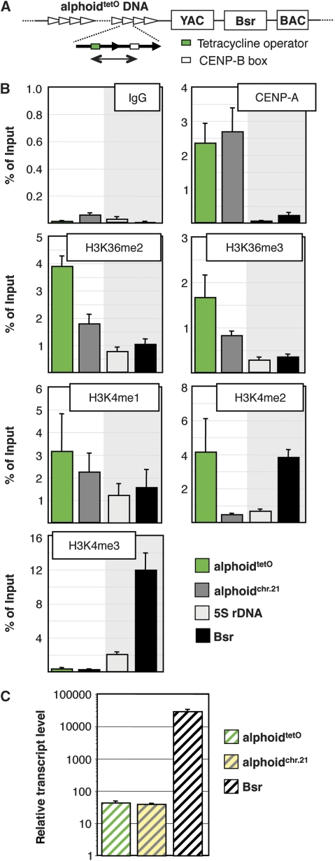
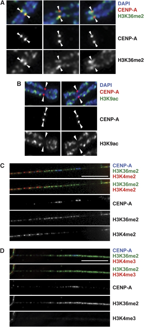
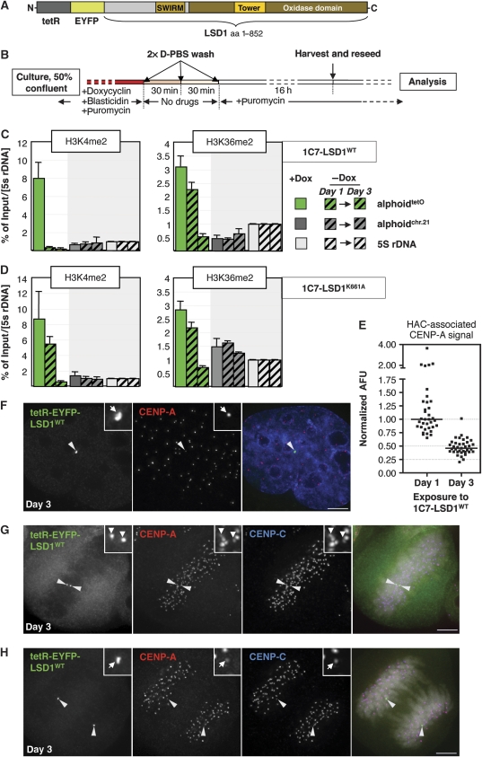
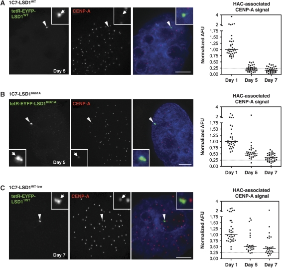
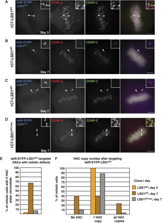
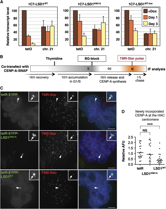
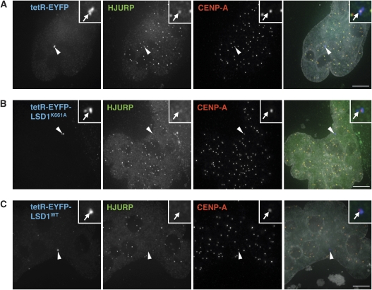
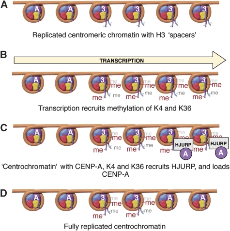
Kinetochores assemble on distinct 'centrochromatin' containing the histone H3 variant CENP-A and interspersed nucleosomes dimethylated on H3K4 (H3K4me2). Little is known about how the chromatin environment at active centromeres governs centromeric structure and function. Here, we report that centrochromatin resembles K4-K36 domains found in the body of some actively transcribed housekeeping genes. By tethering the lysine-specific demethylase 1 (LSD1), we specifically depleted H3K4me2, a modification thought to have a role in transcriptional memory, from the kinetochore of a synthetic human artificial chromosome (HAC). H3K4me2 depletion caused kinetochores to suffer a rapid loss of transcription of the underlying α-satellite DNA and to no longer efficiently recruit HJURP, the CENP-A chaperone. Kinetochores depleted of H3K4me2 remained functional in the short term, but were defective in incorporation of CENP-A, and were gradually inactivated. Our data provide a functional link between the centromeric chromatin, α-satellite transcription, maintenance of CENP-A levels and kinetochore stability.


Conflict of interest statement

The authors declare that they have no conflict of interest.

Figures

Figure 1
Figure 1
Active centromere chromatin displays the signature of elongating RNA polymerase. (A) Schematic of the HAC, derived from Nakano et al (2008), indicating the synthetic alphoidtetO array (green: tetO; white: CENP-B box) and the HAC vector with YAC and BAC cassettes and the blasticidin (Bsr) resistance marker. The region of the alphoidtetO array analysed by ChIP is indicated by green line. (B) ChIP analysis in AB2.2.18.21 cells using antibodies of the indicated reactivity. The synthetic (alphoidtetO) centromere, endogenous chromosome 21 α21-I satellite DNA (alphoidchr.21), the 5S rDNA loci and the Bsr gene on the HAC vector were assessed. Data represent the mean and s.d. of three independent ChIP experiments. Note the different scaling of individual panels reflecting different efficiencies of individual antibodies. (C) Real-time RT–PCR analysis of synthetic HAC centromere (alphoidtetO), actively transcribed Bsr marker and endogenous chromosome 21 satellite (alphoidchr.21). Expression data are normalized to the copy number of the genomic regions and β-actin mRNA levels (see Materials and methods) and displayed as arbitrary numbers. Data represent the mean and s.e.m. of three independent experiments. Note the log scale.
Figure 2
Figure 2
H3K36me2 forms part of the CENP-A chromatin domain. (AD) Immunofluorescence (IF) analysis of unfixed HT1080 chromosomes (A, B) and kinetochore fibres (C, D) using antibodies against CENP-A and the indicated histone modifications. Scale bars: 5 μm.
Figure 3
Figure 3
Centromeric H3K4me2 is not directly required for immediate kinetochore structure or function. (A) Schematic drawing of the tetR-EYFP-LSD1 fusion construct used in transient transfections and for the generation of stable 1C7-LSD1 cell lines. (B) Schematic diagram of the protocol used to wash out doxycycline to allow binding of the tetR fusion construct to the alphoidtetO array. (C, D) ChIP analysis of 1C7 cells stably expressing the wild-type LSD1 (1C7-LSD1WT, C) or the catalytically inactive LSD1K661A (D) tetR fusion constructs prior to, 1 and 3 days after washing out of doxycycline (Dox) to allow binding to the HAC centromere. Targeting of LSD1K661A reduces H3K4me2 levels at the HAC centromere only insignificantly (P=0.16, t-test) within 24 h. Both constructs cause a gradual decrease in centromeric H3K36me2 levels consistent with their ability to repress local transcription (see main text and Figure 6). Two to four independent ChIP experiments where carried out. Percentage of input values for each time point were normalized to the 5S rDNA locus. Error bars represent s.e.m. (E) Quantification of HAC-associated CENP-A staining in 1C7-LSD1WT interphase cells at the indicated time points after washing out of doxycycline. A full time course of this experiment is reproduced in Supplementary Figure S3. Values were normalized to the median value of ‘day 1'. Solid bars indicate the median. (FH) IF analysis of 1C7-LSD1WT cells 3 days after washing out Dox. Interphase CENP-A staining (F) associated with the targeted HAC centromere (arrowheads) remains compacted. At this time point, the HAC sister kinetochores remain structurally and functionally intact as judged by staining for CENP-A and -C in metaphase (G) and anaphase (H). Scale bars: 5 μm.
Figure 4
Figure 4
LSD1 tethering interferes with the maintenance of CENP-A levels at the HAC. (AC) CENP-A IF analysis of interphase 1C7 cells expressing (A) tetR-EYFP-LSD1WT, (B) a catalytically inactive mutant fusion, LSD1K661A or (C) a seven-fold lower level of tetR-EYFP-LSD1WT 5 days after targeting the respective construct to the HAC centromere. Displayed cells represent the median arbitrary fluorescence unit (AFU) of the quantifications shown to the right. Arrowheads depict the HAC. For (C) a ‘day 7' median cell is shown. Scale bars: 5 μm. (Right panels) CENP-A IF signal quantification at HACs targeted by the indicated fusion constructs at the given time points after washing out of Dox. AFU values were normalized to the median value of the ‘day 1' time point. The dotted line indicates the 25% signal mark for orientation. Solid vertical lines represent the median. For 1C7-LSD1WT cells, the corresponding full time course (days 1, 3, 5, 7) quantifications are shown in Supplementary Figure S3B, and day 1 and day 3 values are reproduced in Figure 3E.
Figure 5
Figure 5
LSD1 activity at the HAC centromere correlates with loss of kinetochore function. (AC) IF analysis of metaphase 1C7-LSD1WT cells expressing the fusion construct at high levels at 3 (A) and 7 days (B, C) after Dox washout. Cells were stained for CENP-A (red) and -C (green). Arrowheads indicate the HAC, also shown in the insets. Arrowheads in (A) indicate HAC sister kinetochores under tension. Arrowheads in (B, C) point to unaligned HACs. Arrow in (C) indicates extra HAC. Scale bars: 5 μm. (D) IF analysis as in (A) shows a metaphase 1C7-LSD1WT-low cell 7 days after Dox washout. (E) Analysis of the indicated 1C7-LSD1 clonal lines at the given time points. Late prometa-, meta- and anaphase cells with two detectable HAC sister chromatids (n=36, n=15 and n=29 for clones LSD1WT/day 3, LSD1WT/day 7 and LSD1WT-low/day 7, respectively) were scored. HAC-specific mitotic defects where considered as apparently unaligned HACs in late prometa- and metaphases (see e.g. B, C), as well as mis-segregating HAC sister chromatids in anaphase. (F) Quantification of HAC copy numbers as determined by the EYFP spot pairs in 1C7-LSD1WT clonal cells (n=36, n=39 and n=33 for clones LSD1WT/day 3, LSD1WT/day 7 and LSD1WY-low/day 7, respectively).
Figure 6
Figure 6
Tethering of LSD1 reduces centromeric transcription and impairs the incorporation of newly synthesized CENP-A into the HAC centromere. (A) Real-time RT–PCR analysis of HAC (tetO) and chromosome 21 (chr.21) centromere transcripts in 1C7 cells stably expressing the indicated fusion constructs (WT versus K661A) and the LSD1WT fusion at different levels (high versus low) before, 1 and 3 days after washing out Dox. Differences in the repressive activity of wild-type (high) and mutant LSD1 fusions is statistically significant at both time points (P=0.015 and P<0.001 for day 1 and day 3, respectively; t-test). Data represent the means and s.e.m. of at least three independent experiments. (B) Schematic workflow to determine incorporation of newly synthesized CENP-A into centromeres. (C) 1C7 cells co-transfected with tetR-EYFP, tetR-EYFP-LSD1WT or tetR-EYFP-LSD1K661A and a construct expressing SNAP-tagged CENP-A were analysed by fluorescence microscopy after labelling newly incorporated CENP-A molecules. (D) Quantification of TMR-Star fluorescence signal levels associated with the HAC normalized to the average signal at endogenous centromeres. Asterisks indicate a significant difference, with P<0.001 (t-test).
Figure 7
Figure 7
LSD1 perturbs HJURP localization to the HAC centromere. 1C7 cells were co-transfected with plasmids expressing (A) tetR-EYFP, (B) tetR-EYFP-LSD1WT or (C) tetR-EYFP-LSD1K661A plus a C-terminally LAP-tagged (mRFP) HJURP fusion. Cells displaying centromeric HJURP localization (corresponding to G1 phase) were imaged 2 days after transfection. Arrowheads indicate the HAC. Scale bars: 5 μm.
Figure 8
Figure 8
One model explaining the role of transcription and centromere chromatin in kinetochore maintenance. (A) Following DNA replication, histone H3 (italic ‘3') is inserted in place of CENP-A (‘A'). (B) Transcription through the alphoid array results in methylation of H3 on lysines (4 and 36). (C) During mitotic exit, the ‘centrochromatin' environment, with CENP-A, H3K4me2 and H3K36me2 recruits HJURP, which inserts CENP-A in place of some of the H3 molecules, maintaining the identity of the centromeric chromatin in the course of ongoing cell divisions (D). We have shown that removal of H3K4me2 both lowers centromeric transcription and inhibits the targeting of HJURP to centromere chromatin. Nucleosome drawings by Graham T Johnson.

Comment in

References

    1. Allshire RC, Karpen GH (2008) Epigenetic regulation of centromeric chromatin: old dogs, new tricks? Nat Rev Genet 9: 923–937 - PMC - PubMed
    1. Alonso A, Fritz B, Hasson D, Abrusan G, Cheung F, Yoda K, Radlwimmer B, Ladurner AG, Warburton PE (2007) Co-localization of CENP-C and CENP-H to discontinuous domains of CENP-A chromatin at human neocentromeres. Genome Biol 8: R148. - PMC - PubMed
    1. Barski A, Cuddapah S, Cui K, Roh TY, Schones DE, Wang Z, Wei G, Chepelev I, Zhao K (2007) High-resolution profiling of histone methylations in the human genome. Cell 129: 823–837 - PubMed
    1. Blower MD, Sullivan BA, Karpen GH (2002) Conserved organization of centromeric chromatin in flies and humans. Dev Cell 2: 319–330 - PMC - PubMed
    1. Bouzinba-Segard H, Guais A, Francastel C (2006) Accumulation of small murine minor satellite transcripts leads to impaired centromeric architecture and function. Proc Natl Acad Sci USA 103: 8709–8714 - PMC - PubMed

Publication types

MeSH terms