. 2010 Nov;14(11):2641-5.
doi: 10.1111/j.1582-4934.2010.01186.x.

Telocytes accompanying cardiomyocyte in primary culture: two- and three-dimensional culture environment

Affiliations

Telocytes accompanying cardiomyocyte in primary culture: two- and three-dimensional culture environment

Jin Zhou et al. J Cell Mol Med. 2010 Nov.

Abstract

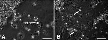
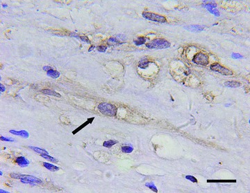
Recently, the presence of telocytes was demonstrated in human and mammalian tissues and organs (digestive and extra-digestive organs, genitourinary organs, heart, placenta, lungs, pleura, striated muscle). Noteworthy, telocytes seem to play a significant role in the normal function and regeneration of myocardium. By cultures of telocytes in two- and three-dimensional environment we aimed to study the typical morphological features as well as functionality of telocytes, which will provide important support to understand their in vivo roles. Neonatal rat cardiomyocytes were isolated and cultured as seeding cells in vitro in two-dimensional environment. Furthermore, engineered myocardium tissue was constructed from isolated cells in three-dimensional collagen/Matrigel scaffolds. The identification of telocytes was performed by using histological and immunohistochemical methods. The results showed that typical telocytes are distributed among cardiomyocytes, connecting them by long telopodes. Telocytes have a typical fusiform cell body with two or three long moniliform telopodes, as main characteristics. The vital methylene blue staining showed the existence of telocytes in primary culture. Immunohistochemistry demonstrated that some c-kit or CD34 immuno-positive cells in engineered heart tissue had the morphology of telocytes, with a typical fusiform cell body and long moniliform telopodes. Also, a significant number of vimentin+ telocytes were present within engineered heart tissue. We suggest that the model of three-dimensional engineered heart tissue could be useful for the ongoing research on the functional relationships of telocytes with cardiomyocytes. Because the heart has the necessary potential of changing the muscle and non-muscle cells during the lifetime, telocytes might play an active role in the heart regeneration process. Moreover, telocytes might be a useful tool for cardiac tissue engineering.


Figures

Fig 1
Fig 1
Telocytes in primary culture at day 7. (A) Typical telocytes with fusiform cell body and two long prolongations (telopodes). (B) Telocytes with two or three prolongations were different from cardiac cells. Bar = 50 μm.
Fig 2
Fig 2
Telocytes in primary culture at day 7. (A) Extremely long and thin prolongations – telopodes (arrow) extended from the body of telocytes. (B) Typical moniliform prolongations (arrows): alternation of thin segments – podomeres – and dilated segments – podoms. Bar = 50 μm.
Fig 3
Fig 3
Telocytes in primary culture at day 7. (A) The prolongation of telocytes ended on cardiomyocytes. (B) Telocytes connected to cardiomyocytes with long prolongations. Bar = 50 μm.
Fig 4
Fig 4
Telocytes in primary culture at day 7. (A) Telocyte around the beating cardiomyocytes. (B) Telocytes (arrows) connected the cardiomyocytes to form networks. Bar = 50 μm.
Fig 5
Fig 5
Methylene blue staining of cells in primary culture at day 10. (A) Telocytes (arrow) with long and extremely thin prolongations, telopodes. (B) Telocytes (arrows) involved in the consistent beating ‘syncytium’. Bar = 50 μm.
Fig 6
Fig 6
Haematoxylin and eosin staining of engineered heart tissue at day 14. (A) Cardiac myocytes with rod-shaped body and nuclei centrally located and elongated oriented longitudinally. (B) Telocytes with two or three long cell processes – telopodes. (C) Telocytes with three long cell processes – telopodes. Arrows indicate dense nodular or vacuolar structures along the prolongation. Bar = 200 μm (A), 20 μm (B, C).
Fig 7
Fig 7
Immunohistochemical staining for c-kit+ cells in engineered heart at day 14. Arrow shows a c-kit+ telocyte in engineered heart tissue. Bar = 20 μm.
Fig 8
Fig 8
Immunohistochemical staining for CD34+ cells in engineered heart at day 14. Arrow shows a CD34+ telocyte in engineered heart tissue. Bar = 20 μm.
Fig 9
Fig 9
Immunohistochemical staining for vimentin in engineered heart at day 14. (A) Vimentin+ cells in engineered heart tissues. (B) Vimentin+ telocyte with long and extremely thin prolongation (telopodes) and vimentin, discontinuously expressed along the prolongation (arrow). Bar = 50 μm (A), 20 μm (B).

References

    1. Popescu LM, Faussone-Pellegrini MS. Telocytes – a case of serendipity: the winding way from interstitial cells of Cajal (ICC), via interstitial Cajal-like cells (ICLC) to Telocytes. J Cell Mol Med. 2010;14:729–40. - PMC - PubMed
    1. Suciu L, Nicolescu MI, Popescu LM. Cardiac telocytes: serial dynamic images in cell culture. J Cell Mol Med. 2010 Doi: . - DOI - PMC - PubMed
    1. Popescu LM, Gherghiceanu M, Kostin S. Telocytes and heart renewing. In: Wang P, Kuo C-H, Takeda N, Singal PK, et al., editors. Adaptation biology and medicine. New Delhi, India: Narosa Publishing House Pvt. Ltd; 2010. pp. 729–2951.
    1. Faussone-Pellegrini MS, Bani D. Relationships between telocytes and cardiomyocytes during pre- and post-natal life. J Cell Mol Med. 2010;14:1061–3. - PMC - PubMed
    1. Kostin S. Myocardial telocytes: a specific new cellular entity. J Cell Mol Med. 2010;14:1917–21. - PMC - PubMed

Publication types