. 2010 Nov;37(11):5896-907.
doi: 10.1118/1.3490556.

Optimization of a dual-energy contrast-enhanced technique for a photon-counting digital breast tomosynthesis system: I. A theoretical model

Affiliations

Optimization of a dual-energy contrast-enhanced technique for a photon-counting digital breast tomosynthesis system: I. A theoretical model

Ann-Katherine Carton et al. Med Phys. 2010 Nov.

Abstract

Purpose: Dual-energy (DE) iodine contrast-enhanced x-ray imaging of the breast has been shown to identify cancers that would otherwise be mammographically occult. In this article, theoretical modeling was performed to obtain optimally enhanced iodine images for a photon-counting digital breast tomosynthesis (DBT) system using a DE acquisition technique.

Methods: In the system examined, the breast is scanned with a multislit prepatient collimator aligned with a multidetector camera. Each detector collects a projection image at a unique angle during the scan. Low-energy (LE) and high-energy (HE) projection images are acquired simultaneously in a single scan by covering alternate collimator slits with Sn and Cu filters, respectively. Sn filters ranging from 0.08 to 0.22 mm thickness and Cu filters from 0.11 to 0.27 mm thickness were investigated. A tube voltage of 49 kV was selected. Tomographic images, hereafter referred to as DBT images, were reconstructed using a shift-and-add algorithm. Iodine-enhanced DBT images were acquired by performing a weighted logarithmic subtraction of the HE and LE DBT images, The DE technique was evaluated for 20-80 mm thick breasts. Weighting factors, w(t) that optimally cancel breast tissue were computed. Signal-difference-to-noise ratios (SDNRs) between iodine-enhanced and nonenhanced breast tissue normalized to the square root of the mean glandular dose (MGD) were computed as a function of the fraction of the MGD allocated to the HE images. Peak SDNR/ mean square root of MGD and optimal dose allocations were identified. SDNR/ mean square root of MGD and dose allocations were computed for several practical feasible system configurations (i.e., determined by the number of collimator slits covered by Sn and Cu). A practicalsystem configuration an d Sn-Cu filterpair that accounts for the trade-off between SDNR, tube-output, and MGD were selected.

Results: w(t) depends on the Sn-Cu filter combination used, as well as on the breast thickness; to optimally cancel 0% with 50% glandular breast tissue, w(t) values were found to range from 0.46 to 0.72 for all breast thicknesses and Sn-Cu filter pairs studied. The optimal w(t) values needed to cancel all possible breast tissue glandularites vary by less than 1% for 20 mm thick breasts and 18% for 80 mm breasts. The system configuration where one collimator slit covered by Sn is alternated with two collimator slits covered by Cu delivers SDNR/ mean square root of MGD nearest to the peak value. A reasonable compromise is a 0.16 mm Sn-0.23 mm Cu filter pair, resulting in SDNR values between 1.64 and 0.61 and MGD between 0.70 and 0.53 mGy for 20-80 mm thick breasts at the maximum tube current.

Conclusions: A DE acquisition technique for a photon-counting DBT imaging system has been developed and optimized.


Figures

Figure 1
Figure 1
Design principle of the photon-counting XC Mammo-3T (XCounter AB, Danderyd, Sweden). The x-ray tube, prepatient collimator, and camera (consisting of 48 linear detectors) are mounted on an E-arm. The prepatient collimator is used to define 48 fan-shaped beams; each is precisely aligned with the x-ray tube and a detector. Images are produced by linearly scanning the E-arm past the breast; in the process, 48 images of the breast are produced, each at a unique angle. The dual-energy implementation was obtained by differentially filtering the 48 fan beams by Sn and Cu. In this way, LE and HE images are obtained simultaneously in a single scan. In the shown configuration, one Sn filter is alternated by one Cu filter. As such, 24 detectors capture a LE image and 24 detectors capture a HE image. Other system configurations are obtained by alternately filtering a different number of fan beams with Sn or Cu.
Figure 2
Figure 2
Illustration of a Sn (LE) and Cu (HE) en x-ray spectrum detected by the XC-Mammo 3T XCounter detector after transmission through a 20 mm thick breast.
Figure 3
Figure 3
Air kerma per mAs at 58 cm from the focus of the x-ray source as a function of Sn and Cu filter thickness.
Figure 4
Figure 4
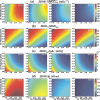
(a) wt giving optimal breast tissue cancellation for (fg1,fg2)=(0.0,0.5). (b) Average energy difference between detected Sn and Cu spectra. (c) Difference (%) between wt values that optimally cancel (fg1,fg2)=(0.0,0.5) and (fg1,fg2)=(0.5,1.0). All results are shown as a function of Sn–Cu filter pair and breast thickness (indicated in white in the upper right corners).
Figure 5
Figure 5
wt giving optimal breast tissue cancellation as a function of (fg1,fg2) using a 0.16 mm Sn–0.23 mm Cu filter pair. Results are shown for breast thicknesses of 20, 40, 60, and 80 mm.
Figure 6
Figure 6
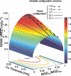
SDNRMGDTotal between 1 mg∕cm2 iodine-enhanced and nonenhanced 40 mm thick breast tissue as a function of MGDCu∕MGDTotal and Cu filter thickness. A 0.16 mm Sn filter was used. The lines drawn on the surface indicate SDNRMGDTotal for various fixed system configurations.
Figure 7
Figure 7
(a) SDNRmaxMGDTotal between 1 mg∕cm2 iodine-enhanced and nonenhanced breast tissue, (b) optimal MGDCu∕MGDTotal, and (c) collimator ratios for the variable configuration as a function of Sn–Cu filter pair and breast thickness (indicated in the upper right corner). The calculations were performed for breasts with glandular tissue fraction fg=0.5.
Figure 8
Figure 8
(a) SDNRMGDTotal between 1 mg∕cm2 iodine-enhanced and nonenhanced breast tissue, (b) MGDCu∕MGDTotal, (c) MGDTotal∕mA s, and (d) SDNR at 140 mA as a function of Sn–Cu filter pair and breast thickness for the 1:2 fixed system configuration. The calculations were performed for breasts with glandular tissue fraction fg=0.5. In (a), the region where SDNRMGDTotal exceeds 85% of SDNRmaxMGDTotal occurs between the dotted lines for 20 and 40 mm thick breasts and below the dotted lines for 60 and 80 mm thick breast.
Figure 9
Figure 9
(a) Principle of resampling when using the shift-and-add algorithm. Projection n (1) and projection n (2) are projection images acquired at two different angles. The colored pixels represent the shadow of an unknown object. To reconstruct the image with the object in focus, Projection n is shifted with respect to Projection n to have the shadows registered (3). Projection n is then resampled to have the pixels of Projection n and Projection n registered (4). (b) Standard deviation in the SI of the resampled projection image as a function of x, the resampling factor.

References

    1. Weidner N., Semple J. P., Welch W. R., and Folkman J., “Tumor angiogenesis and metastasis: Correlation in invasive breast carcinoma,” N. Engl. J. Med. NEJMAG 324, 1–8 (1991).10.1056/NEJM199101033240101 - DOI - PubMed
    1. Hylton N. M., “Vascularity assessment of breast lesions with gadolinium-enhanced MR imaging,” Magn. Reson Imaging Clin. N. Am. ZZZZZZ 9, 321–331 (2001). - PubMed
    1. Kuhl C. K., Schild H. H., and Morakkabati N., “Dynamic bilateral contrast-enhanced MR imaging of the breast: Trade-off between spatial and temporal resolution,” Radiology RADLAX 263, 789–800 (2005).10.1148/radiol.2363040811 - DOI - PubMed
    1. Schnall M. D. et al., “Diagnostic architectural and dynamic features at breast MR imaging: Multicenter study,” Radiology RADLAX 238, 42–53 (2006).10.1148/radiol.2381042117 - DOI - PubMed
    1. Saslow D. et al. “American Cancer Society guidelines for breast screening with MRI as an adjunct to mammography,” Ca-Cancer J. Clin. CAMCAM 57, 75–89 (2007).10.3322/canjclin.57.2.75 - DOI - PubMed

Publication types