. 2011 Jan 13;115(1):158-67.
doi: 10.1021/jp107577k. Epub 2010 Dec 15.

Morphological changes induced by the action of antimicrobial peptides on supported lipid bilayers

Affiliations

Morphological changes induced by the action of antimicrobial peptides on supported lipid bilayers

Ahmad Arouri et al. J Phys Chem B. .

Abstract

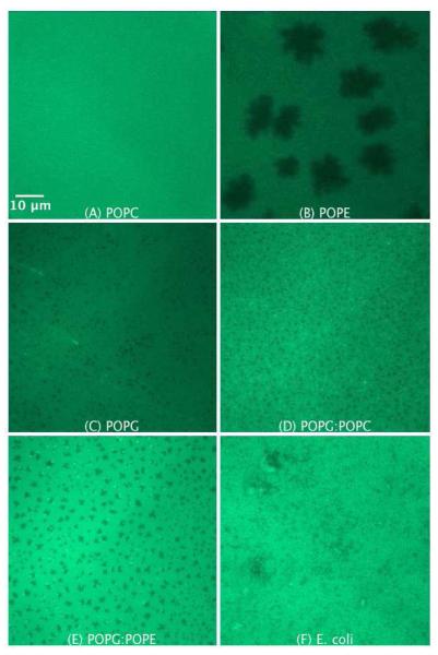
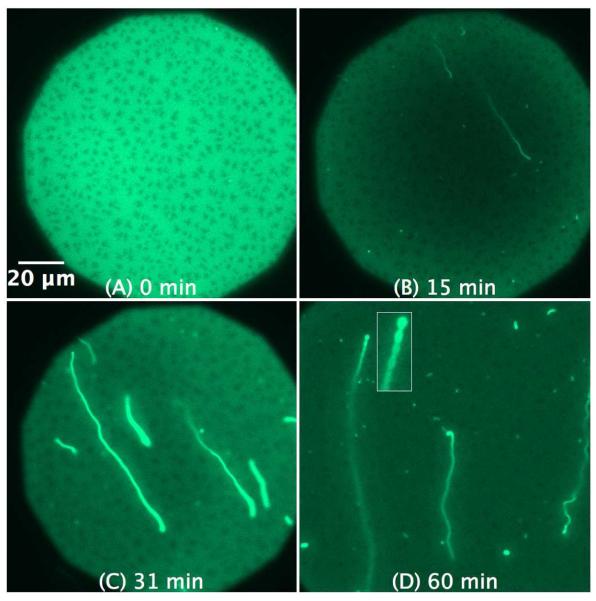
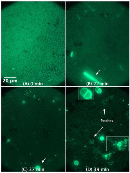
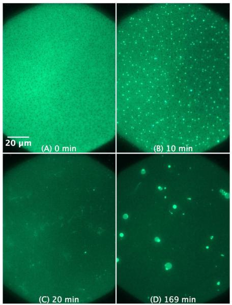
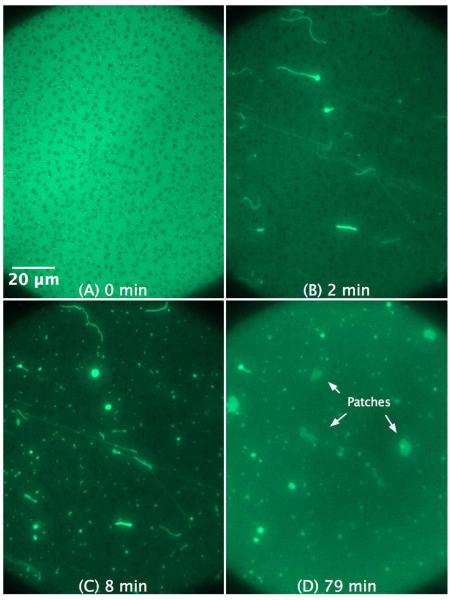
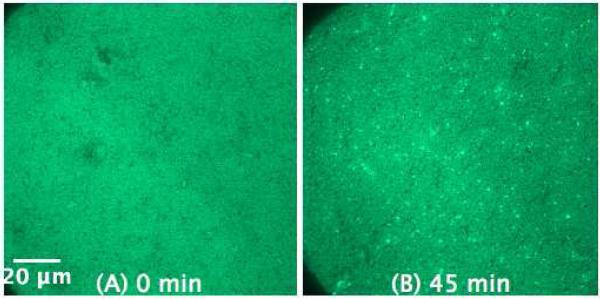
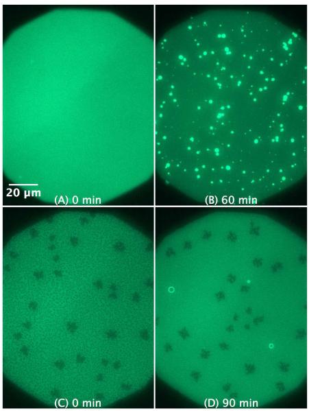
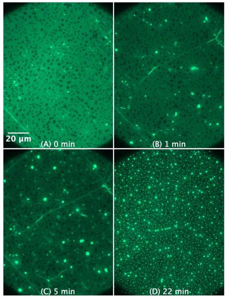
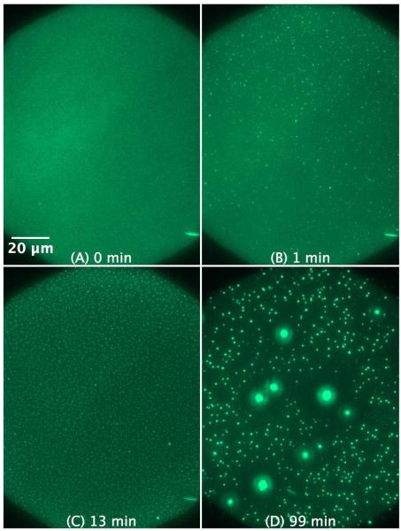
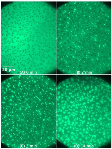
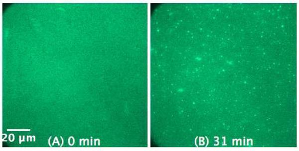
We utilized epifluorescence microscopy to investigate the morphological changes in labeled lipid bilayers supported on quartz surfaces (SLBs) induced by the interaction of cationic antimicrobial peptides with the lipid membranes. The SLBs were prepared from 1-palmitoyl-2-oleoyl-sn-glycero-3-phosphoglycerol (POPG), 1-palmitoyl-2-oleoyl-sn-glycero-3-phosphocholine (POPC), 1-palmitoyl-2-oleoyl-sn-glycero-3-phosphoethanolamine (POPE) and mixtures thereof as well as from Escherichia coli lipid extract. We succeeded in the preparation of POPG and POPG-rich SLBs without the necessity to use fusogenic agents such as calcium by using the Langmuir-Blodgett/Langmuir-Schaefer transfer method. The adsorption of the peptides to the SLBs was initially driven by electrostatic interactions with the PG headgroups and led to the formation of lipid protrusions bulging out from the lipid layer facing the bulk, originating particularly from domain boundaries and membrane defects. The shape, size, and frequency of the lipid protrusions are mainly controlled by the peptide macroscopic properties and the membrane composition. A restructuring of the lipid protrusions into other structures can also occur over time.


Figures

Figure 1
Figure 1
An illustration showing the amphipathic structure of the helical KLA1 (A and B) and of the cyclic hexapeptide C-RW (C and D), where the basic amino acids are oriented in one direction and the hydrophobic residues are aligned in the opposite direction. In the side view (A and C) as well as the Connolly surface (B and D), lys (K) is coloured blue, arg (R) is coloured dark blue, leu (L) is coloured green, ala (A) is coloured grey, trp (W) is coloured pink, phe (F) is coloured sky blue, and the backbone is coloured light gray.
Figure 2
Figure 2
SLB prepared by LB/LS transfer of the pure lipids POPC (A), POPE (B), POPG (C), POPG/POPC 1:1 (D), POPG/POPE 1:1 (E), and E. coli lipid extract (F). The lipids were premixed with 0.75 mol% NBD-DPPE. The dark areas represent dye-depleted domains.
Figure 3
Figure 3
SLB prepared by LB/LS transfer of POPG (NBD-DPPE 0.75 mol%). The images illustrate the effect and morphological changes that occur over time (0 to 60 min) after the addition of 1 μM C-RW into the buffer. The subsequent images became darker as compared to the control image due to the photobleaching effect. The inset in (D) is a magnification of the pearl-chain instability.
Figure 4
Figure 4
SLB prepared by LB/LS transfer of POPG (NBD-DPPE 0.75 mol%). The figures illustrate the effect and morphological changes that occur over time (0 to 39 min) after the addition of 4 μM C-RW into the buffer. The subsequent images became darker as compared to the control image due to the photobleaching effect. The insets in (D) are magnifications of some structures in the image.
Figure 5
Figure 5
SLB prepared by LB/LS transfer of POPG/POPC 1:1 (NBD-DPPE 0.75 mol%). The figures illustrate the effect and morphological changes that occur over time (0 to 169 min) after the addition of 4 μM C-RW into the buffer.
Figure 6
Figure 6
SLB prepared by LB/LS transfer of POPG/POPE 1:1 (NBD-DPPE 0.75 mol%). The figures illustrate the effect and morphological changes that occur over time (0 to 79 min) after the addition of 4 μM C-RW into the buffer.
Figure 7
Figure 7
SLB prepared by LB/LS transfer of E. coli lipid extract (NBD-DPPE 0.75 mol%) before (A) and 45 min after the addition of 1.2 μM C-RW into the buffer (B).
Figure 8
Figure 8
SLB prepared by LB/LS transfer of POPC (left) and POPE (right) (NBD-DPPE 0.75 mol%) before (A and C, respectively) as well as 60 and 90 min after the addition of 4 μM KLA1 into the buffer (B and D, respectively).
Figure 9
Figure 9
SLB prepared by LB/LS transfer of POPG (NBD-DPPE 0.75 mol%). The figures illustrate the effect and morphological changes that occur over time (0 to 22 min) after the addition of 4 μM KLA1 into the buffer. The bright lines in the SLB shown in (B) and (C) are most probably membrane deformations that were initially formed during the peptide injection process.
Figure 10
Figure 10
SLB prepared by LB/LS of POPG/POPC 1:1 (NBD-DPPE 0.75 mol%). The figures illustrate the effect and morphological changes that occur over time (0 to 99 min) after the addition of 4 μM KLA1 into the buffer.
Figure 11
Figure 11
SLB prepared by LB/LS of POPG/POPE 1:1 (NBD-DPPE 0.75 mol%). The figures illustrate the effect and morphological changes that occur over time (0 to 14 min) after the addition of 4 μM KLA1 into the buffer.
Figure 12
Figure 12
SLB prepared by LB/LS of E. coli lipid extract (NBD-DPPE 0.75 mol%) before (A) and 31 min after the addition of 5 μM KLA1 into the buffer (B).

References

    1. Gordon YJ, Romanowski EG, McDermott AM. Curr. Eye Res. 2005;30:505. - PMC - PubMed
    1. Radek K, Gallo R. Semin. Immunopathol. 2007;29:27. - PubMed
    1. Bechinger B, Lohner K. Biochim. Biophys. Acta. 2006;1758:1529. - PubMed
    1. Brogden KA. Nat. Rev. Microbiol. 2005;3:238. - PubMed
    1. Chan DI, Prenner EJ, Vogel HJ. Biochim. Biophys. Acta. 2006;1758:1184. - PubMed

Publication types

MeSH terms

Substances