. 2011 Feb 1;50(4):581-90.
doi: 10.1021/bi101192f. Epub 2010 Dec 30.

Insights into base selectivity from the 1.8 Å resolution structure of an RB69 DNA polymerase ternary complex

Affiliations
Free PMC article

Insights into base selectivity from the 1.8 Å resolution structure of an RB69 DNA polymerase ternary complex

Mina Wang et al. Biochemistry. .
Free PMC article

Abstract

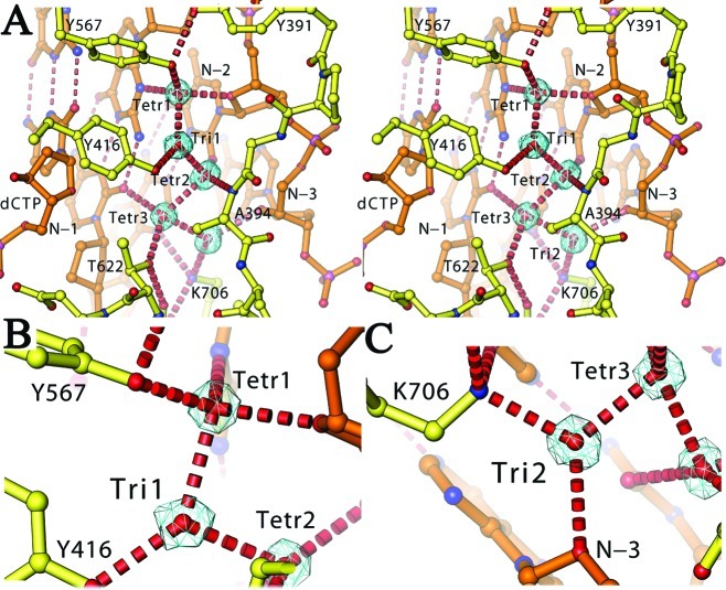
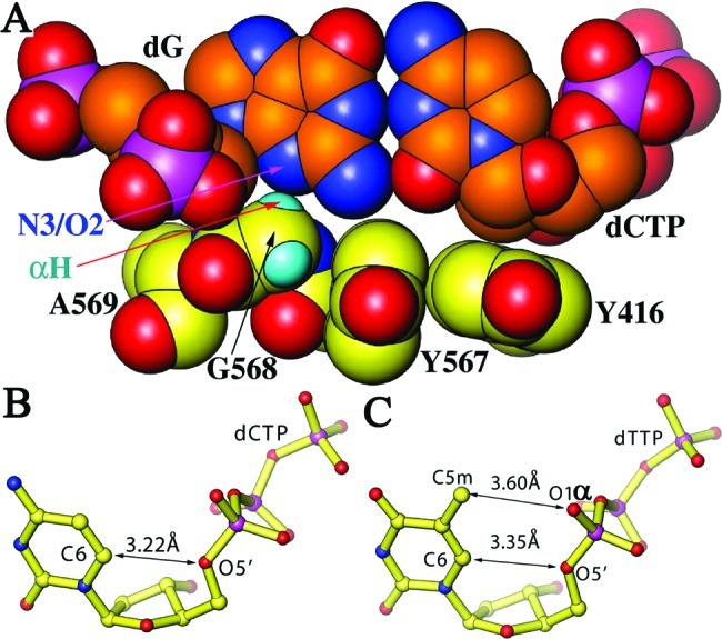
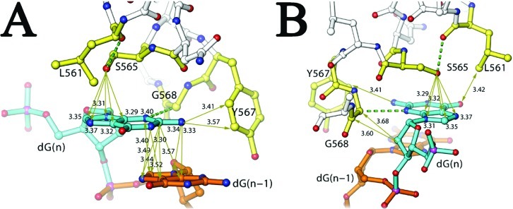
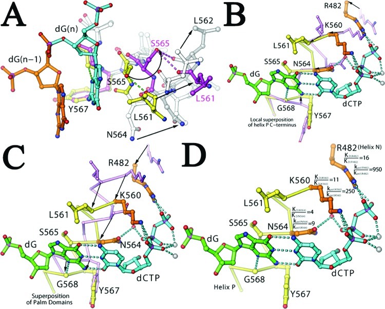
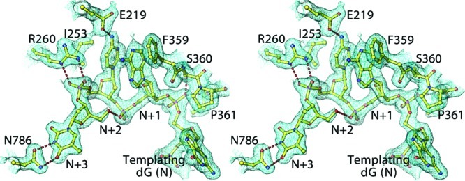
Bacteriophage RB69 DNA polymerase (RB69 pol) has served as a model for investigating how B family polymerases achieve a high level of fidelity during DNA replication. We report here the structure of an RB69 pol ternary complex at 1.8 Å resolution, extending the resolution from our previously reported structure at 2.6 Å [Franklin, M. C., et al. (2001) Cell 105, 657-667]. In the structure presented here, a network of five highly ordered, buried water molecules can be seen to interact with the N3 and O2 atoms in the minor groove of the DNA duplex. This structure reveals how the formation of the closed ternary complex eliminates two ordered water molecules, which are responsible for a kink in helix P in the apo structure. In addition, three pairs of polar-nonpolar interactions have been observed between (i) the Cα hydrogen of G568 and the N3 atom of the dG templating base, (ii) the O5' and C5 atoms of the incoming dCTP, and (iii) the OH group of S565 and the aromatic face of the dG templating base. These interactions are optimized in the dehydrated environment that envelops Watson-Crick nascent base pairs and serve to enhance base selectivity in wild-type RB69 pol.


Figures

Figure 1
Figure 1
Five buried water molecules. (A−C) Standard and close-up views of stereodiagrams for five buried water molecules (contoured at 0.7σ and edited to remove nonsolvent density for the sake of clarity), including two planar triangularly coordinated water molecules (B and C).
Figure 2
Figure 2
Eclipsed conformation of the incoming dCTP in the nascent base pair. (A) Recognition of N3 and O2 hydrogen bond acceptors by the G568 Cα hydrogen (small cyan spheres). (B) Eclipsed conformation of dCTP in this ternary complex. (C) Eclipsed conformation of dTTP in the previously reported RB69 pol ternary complex (6).
Figure 3
Figure 3
Rigidity of the templating nucleotide-binding pocket. (A) Interactions with interatomic distances (in angstroms) indicated for the base of the dG (in cyan) observed in this ternary complex. Essential residues for the binding pocket are colored yellow, and nonessential residues are colored silver. The nucleotide 3′ to the dG is colored gold. (B) Interactions of the ribosyl moiety of dG with G568.
Figure 4
Figure 4
Formation of the NBP upon Fingers domain closing. (A) Superposition of the ternary complex (yellow and silver) with the apo structure (magenta and silver) using main atoms of Y577, G568, and A569 shows the effects of the kinked-to-straight helical transition on interactions with the dG. Large movements of key residues are indicated. (B) Same as panel A, but using Cα atoms of the C-terminus of helix P for superposition. (C) Same as panel A, but using Cα atoms of the Palm domain. (D) Contribution of K560, N564, and R482 (gold) to kinetic parameters for the correct dNTP in the ternary complex is indicated next to each residue as the KD(mutant)/KD(wt) ratio and the kpol(wt)/kpol(wt) ratio when each residue was substituted by an Ala residue (31).
Figure 5
Figure 5
Helices of the Fingers domain. (A) Final 2FoFc map contoured at 1.4σ superimposed with the refined model of helix P. (B) Hydrogen atoms are generated for explicit hydrogen bonds in the backbone of helix P. Nonstandard backbone hydrogen bonds are colored yellow. (C) Superposition of helices of the Fingers domain between the structure of the current complex (yellow) and the previous apo structure (cyan and yellow) of RB69 pol using the Cα atoms of H485−T554 (5,6). Large displacements for G569 and V573 between the two structures are indicated. (D) Same as panel C, but the Cα atoms of L562−L566 were used for superposition. (E) Same as panel C, but the Cα atoms of G568−N572 were used for superposition. (F) Helix P of the apo structure with two water molecules inserted into its backbone. (G) Superposition of helix P (yellow) of our ternary complex with its equivalent helix (cyan) of the ternary complex of ϕ29 pol (16). (H) Superposition of helix P (yellow) of the apo structure of RB69 pol with its equivalent helix (cyan) of the apo form of ϕ29 pol (16).
Figure 6
Figure 6
Structure of the overhanging 5′ template in stereodiagram superimposed onto the final 2FoFc map contoured at 0.5σ.
Figure 7
Figure 7
Primer extension assays of mismatched DNA duplexes. Lanes 1 and 8 were no-extension control experiments. Lanes 2−7 were primer extension assays with all four dNTPs and wild-type RB69 pol (exo) incubated for 15 s and lanes 9−14 for 2 min. Lane 1: P/T1 only, without enzyme or dNTPs. Lane 8: P/T1 and enzyme without dNTPs. Lanes 2−7 and 9−14: P/Ts and enzyme with four dNTPs, where the P/Ts for the six sequential lanes are P/T1, P/T2, P/T3, P/T4, P/T5, and P/T6, respectively. Sequences of P/T1−P/T6 are given in the footnote of Table 2.

References

    1. Echols H.; Goodman M. F. (1991) Fidelity mechanisms in DNA replication. Annu. Rev. Biochem. 60, 477–511. - PubMed
    1. Joyce C. M.; Steitz T. A. (1994) Function and structure relationship in DNA polymerases. Annu. Rev. Biochem. 63, 777–822. - PubMed
    1. Johnson K. A. (2010) The kinetic and chemical mechanism of high-fidelity DNA polymerases. Biochim. Biophys. Acta 1804, 1041–1048. - PMC - PubMed
    1. Braithwaite D. K.; Ito J. (1993) Compilation, alignment, and phlogenetic relationships of DNA polymerases. Nucleic Acids Res. 21, 787–802. - PMC - PubMed
    1. Wang J.; Sattar A. K.; Wang C. C.; Karam J. D.; Konigsberg W. H.; Steitz T. A. (1997) Crystal structure of a pol α family replication DNA polymerase from bacteriophage RB69. Cell 89, 1087–1099. - PubMed

Publication types