Review
. 2011 Jan 1;433(1):31-42.
doi: 10.1042/BJ20100985.

E2s: structurally economical and functionally replete

Affiliations
Review

E2s: structurally economical and functionally replete

Dawn M Wenzel et al. Biochem J. .

Erratum in

  • Biochem J. 2011 Jan 14;433(3):535

Abstract

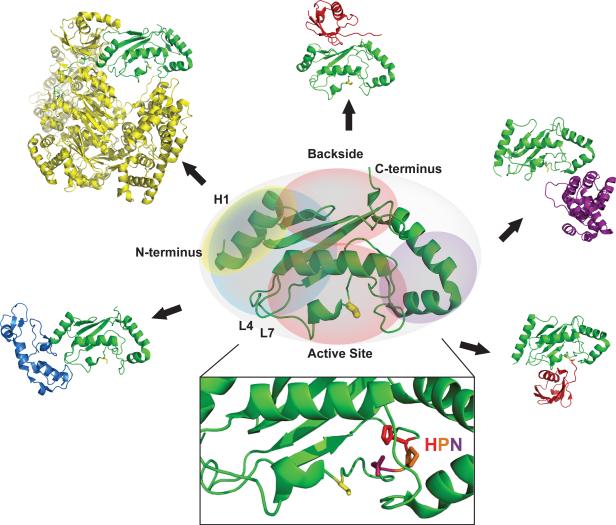
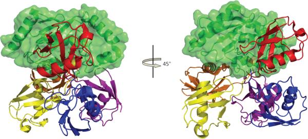
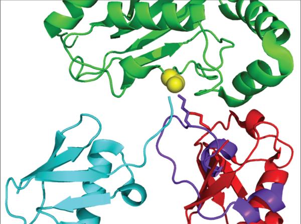
Ubiquitination is a post-translational modification pathway involved in myriad cellular regulation and disease pathways. The Ub (ubiquitin) transfer cascade requires three enzyme activities: a Ub-activating (E1) enzyme, a Ub-conjugating (E2) enzyme, and a Ub ligase (E3). Because the E2 is responsible both for E3 selection and substrate modification, E2s function at the heart of the Ub transfer pathway and are responsible for much of the diversity of Ub cellular signalling. There are currently over 90 three-dimensional structures for E2s, both alone and in complex with protein binding partners, providing a wealth of information regarding how E2s are recognized by a wide variety of proteins. In the present review, we describe the prototypical E2-E3 interface and discuss limitations of current methods to identify cognate E2-E3 partners. We present non-canonical E2-protein interactions and highlight the economy of E2s in their ability to facilitate many protein-protein interactions at nearly every surface on their relatively small and compact catalytic domain. Lastly, we compare the structures of conjugated E2~Ub species, their unique protein interactions and the mechanistic insights provided by species that are poised to transfer Ub.


Figures

Figure 1
Figure 1
The 1, 2, 3's of protein ubiquitination. Simplified schematic shows the three enzymatic activities associated with the central paradigm of protein ubiquitination: E1, the Ub-activating enzyme; E2, the Ub-conjugating enzyme; and E3, the Ub ligase. Mechanistically, there are two types of E3s, the RING/UBox-type E3s that effect transfer of Ub directly from the active site of an E2 to a lysine residue of a substrate, and the HECT-type E3s that facilitate Ub transfer from an E2 to substrate via a thioester intermediate on the E3. Auto-ubiquitination of the E3 is also observed in some cases and can be used as a proxy signal to assay the activity of an E2/E3 pair in the absence of a substrate. The best studied fate of poly-ubiquitinated substrates is degradation by the proteasome (not depicted).
Figure 2
Figure 2
Structure of a Ubc domain and its interaction surfaces. Shown in the center is the E2, Ubc13 in ribbon structure (PDB code 2GMI), with interaction surfaces described in the text colored. The E2 in each interacting pair is shown as green. Clockwise from lower left corner: the E1/E3-binding surface as seen in complex of Ubc13 with the RING E3 Traf6 (blue) (PDB code 3HCT) and in Ubc12 in complex with the NEDD8-activating E1 (yellow) (PDB code 2NVU); backside-binding surface as seen in the E2/Ub complex, UbcH5c/Ub (red) (PDB code 2FUH); substrate-binding surface as seen in the SUMO E2 Ubc9 in complex with its substrate RANGAP1(purple) (PDB code 1Z5S); activated Ub/Ubl surface as seen in UbcH5b~Ub complex (PDB code 3A33). The inset shows the highly conserved HPN motif at the active site, with the active site Cys shown as yellow stick representation.
Figure 3
Figure 3
RING- and HECT-type E3s recognize the same surface on the E2 UbcH7. Shown are the archetypes for canonical E2/E3 interactions: A) UbcH7 in complex with the HECT-E3, E6AP (PDB code 1C4Z); B) UbcH7 in complex with the RING-E3, cCbl (PDB code 1FBV).
Figure 4
Figure 4
Ub/Ubl and non-canonical E3 binding to the E2 backside. Top, non-covalent complexes of E2s with Ub/Ubl (red) bound on the backside. Bottom, non-canonical E3 (blue) binding uses the same E2 surface. PDB codes are (left to right from the top: 2EKE, 2UYZ, 2FUH, 3A4S, 3H8K, 1Z5S).
Figure 5
Figure 5
Ub conjugated to an E2 assumes multiple orientations relative to the E2. Shown in green, the E2 Ubc of each structure has been superimposed; active site Cys is shown in yellow spheres. Ub moiety in each structure is shown in a different color: Red, Ubc1~Ub (PDB code 1FXT); Blue, UbcH5b~Ub in complex with Nedd4L (PDB code 3JVZ); Yellow, Ubc13~Ub (PDB code 2GMI); Orange, UbcH5b~Ub (PDB code 3A33); Purple, UbcH8~Ub (PDB code 2KJH). View on the left shows the same E2 orientation as in Figures 2, 3, and 4; view on the right is rotated 45 degrees about the vertical axis.
Figure 6
Figure 6
Structure of an E3/E2~Ub complex: HECT-E3, Nedd4L (blue), in complex with UbcH5b~Ub (green/red). Left, HECT N-lobe contacts E1/E3 surface on UbcH5, as observed in previous structures (see Figure 3) and HECT C-lobe makes contacts with the ~Ub moiety. The NEDD4L active site is shown in yellow (PDB code 3JVZ). Right, view rotated by 180 degrees to show NEDD4L residues analogous to those observed in Rsp5 and SMURF2 that are important for non-covalent Ub binding in the N-lobe, shown as orange spheres.
Figure 7
Figure 7
Position of substrate lysine approach into the E2 active site. Lys63 of a putative substrate Ub in the formation of a Lys63-linked Ub chain observed in a structure of Ubc13~Ub/Mms2 (PDB code 2GMI) is shown in red stick representation. Substrate lysine residue involved in isopeptide linkage to Ubl (SUMO) observed in a structure of E2/E3/product (Ubc9/Nup358/SUMO-RanGAP1; PDB code 1Z5S) is shown in purple stick representation. The E2s of each structure were superimposed for this figure and shown in green, with active site Cys in yellow spheres (conjugated Ub in Ubc13~Ub is shown in cyan).

References

    1. Hershko A, Heller H, Elias S, Ciechanover A. Components of ubiquitin-protein ligase system. J. Biol. Chem. 1983;258:8206–8214. - PubMed
    1. Christensen DE, Brzovic PS, Klevit RE. E2-BRCA1 RING interactions dictate synthesis of mono- or specific polyubiquitin chain linkages. Nat. Struct. Mol. Biol. 2007;14:941–948. - PubMed
    1. Roderigo-Brenni MC, Morgan DO. Sequential E2s drive polyubiquitin chain assembly on APC targets. Cell. 2007;130:127–39. - PubMed
    1. Windheim M, Peggie M, Cohen P. Two different classes of E2 ubiquitinconjugating enzymes are required for the mono-ubiquitination of proteins and elongation by polyubiquitin chains with a specific topology. Biochem J. 2008;409:723–9. - PubMed
    1. Tang Z, Hecker CM, Scheschonka A, Betz H. Protein interactions in the sumoylation cascade: lessons from X-ray structures. FEBS J. 2008;275:3003–15. - PubMed

Publication types

MeSH terms

Substances