Review
. 2011 Jan;13(1):1-14.
doi: 10.1111/j.1477-2574.2010.00225.x. Epub 2010 Nov 15.

An analytical review of vasculobiliary injury in laparoscopic and open cholecystectomy

Affiliations
Review

An analytical review of vasculobiliary injury in laparoscopic and open cholecystectomy

Steven M Strasberg et al. HPB (Oxford). 2011 Jan.

Abstract

Objectives: Biliary injuries are frequently accompanied by vascular injuries, which may worsen the bile duct injury and cause liver ischemia. We performed an analytical review with the aim of defining vasculobiliary injury and setting out the important issues in this area.

Methods: A literature search of relevant terms was performed using OvidSP. Bibliographies of papers were also searched to obtain older literature.

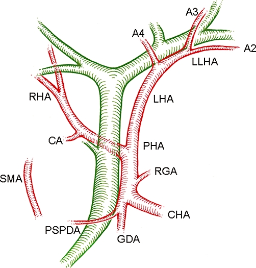
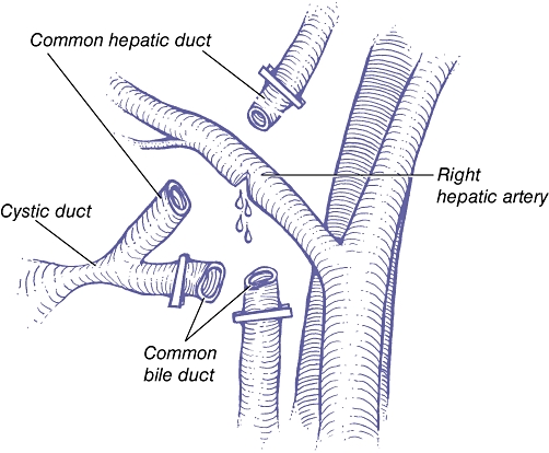
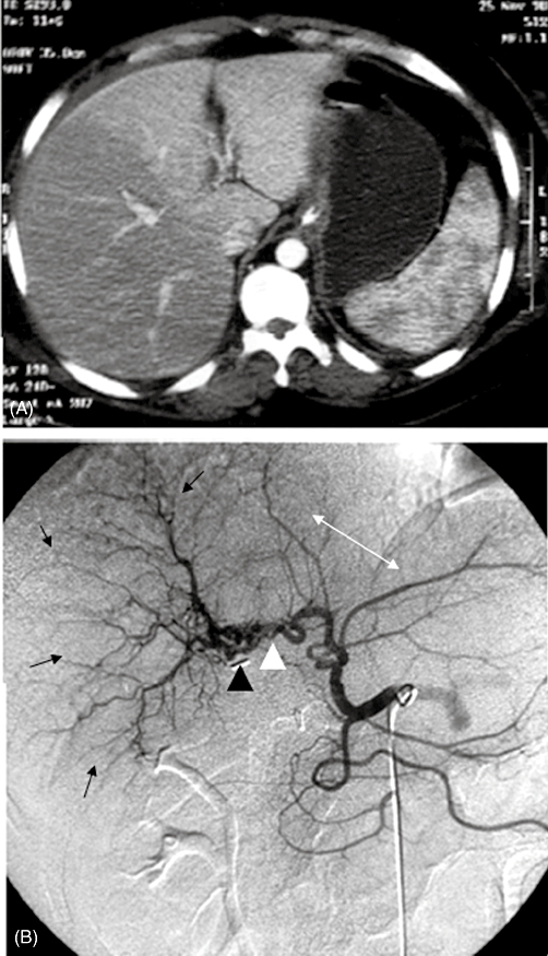
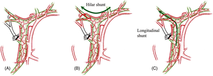
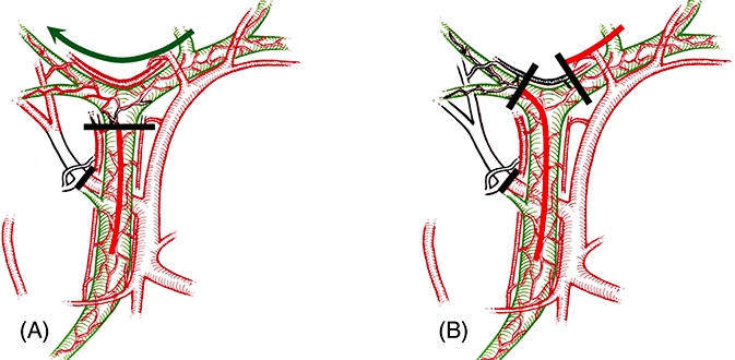
Results: Vasculobiliary injury was defined as: an injury to both a bile duct and a hepatic artery and/or portal vein; the bile duct injury may be caused by operative trauma, be ischaemic in origin or both, and may or may not be accompanied by various degrees of hepatic ischaemia. Right hepatic artery (RHA) vasculobiliary injury (VBI) is the most common variant. Injury to the RHA likely extends the biliary injury to a higher level than the gross observed mechanical injury. VBI results in slow hepatic infarction in about 10% of patients. Repair of the artery is rarely possible and the overall benefit unclear. Injuries involving the portal vein or common or proper hepatic arteries are much less common, but have more serious effects including rapid infarction of the liver.

Conclusions: Routine arteriography is recommended in patients with a biliary injury if early repair is contemplated. Consideration should be given to delaying repair of a biliary injury in patients with occlusion of the RHA. Patients with injuries to the portal vein or proper or common hepatic should be emergently referred to tertiary care centers.


Figures

Figure 1
Figure 1
Blood supply to the bile ducts. Element 1: the supplying arteries. The arteries shown can all twig to the marginal arteries or, in some cases, directly supply the epicholedochal plexus. SMA, superior mesenteric artery; PSPDA, posterior superior pancreatoduodenal artery (the most important and constant artery); CHA, common hepatic artery; PHA, proper hepatic artery; RGA, right gastric artery; GDA, gastroduodenal artery; RHA, right hepatic artery; LHA, left hepatic artery; CA, cystic artery; LLHA, left lateral hepatic artery; A2, A3, A4, arteries to segments 2, 3 and 4. Replaced arteries can also supply the bile ducts
Figure 2
Figure 2
Blood supply to the bile ducts. Element 2: the marginal arteries. Marginal arteries are disposed at 3, 9 and, rarely, 12 o'clock (not shown) on the common bile duct/common hepatic duct. The hilar marginal artery runs across the top of the confluence of the right and left hepatic ducts
Figure 3
Figure 3
Blood supply to the bile ducts. Element 3: the epicholedochal plexus. The epicholedochal plexus is supplied by the marginal arteries and sometimes directly by the supplying arteries. The part of the network around the confluence of the right and left hepatic ducts has been termed the ‘hilar plexus’. However, it is continuous with the plexus that surrounds the common duct and is probably best considered as the hilar component of the epicholedochal plexus
Figure 4
Figure 4
Pathogenesis of the ‘classical’ injury. 1. The common bile duct is mistaken as the cystic duct and is clipped and divided. 2. The dissection is carried up along the left side of the common hepatic duct in the belief that this is the underside of the gallbladder. 3. The common hepatic duct is transected as the surgeon tries to dissect what he or she believes is the gallbladder from the liver bed. If the structure is recognized as a bile duct at this point, it is often thought to be a second cystic duct or an accessory duct. As the common hepatic duct is divided, the right hepatic artery is often injured (see Fig. 6)
Figure 5
Figure 5
The final stage of the ‘classical’ injury. The hepatic duct has been divided. The right hepatic artery, which lies immediately behind the common hepatic duct, is injured. (Redrawn from Davidoff et al.3)
Figure 6
Figure 6
Right hepatic artery (RHA) vasculobiliary injury with collateral flow from left hepatic artery and atrophy of right liver. (A) Computed tomography scan of liver shortly after injury. The arterial phase shows no filling of right liver. (B) Arteriogram performed 2 years later. Abundant arterial collaterals extend from the left hepatic artery to the RHA along the hilar plexus (white arrowhead). The clip which occluded the RHA is also seen (black arrowhead). The arterial pattern of the right liver shows crowding (black arrows) indicative of atrophy of the right liver, whereas the arterial pattern of the left liver shows elongation and spreading characteristic of hypertrophy of the left liver. (Reproduction of original photographs from Mathisen et al. by permission)
Figure 7
Figure 7
Effect on hepatic blood flow of an injury to the right hepatic artery (RHA) without biliary injury. (A) Occlusion of the RHA results in ischaemia of the right liver, but (B, C) flow is restored by preformed collateral arterial shunts
Figure 8
Figure 8
Vasculobiliary injury involving the right hepatic artery (RHA). (A) E1–3 injuries leave the hilar shunt open but obstruct the longitudinal shunt and may induce greater hepatic ischaemia than RHA occlusion only. (B) E4 injuries induce greater ischaemia than right hepatic injuries alone by obstructing the important hilar shunt and the longitudinal shunt
Figure 9
Figure 9
Computed tomography scan showing infarction of right liver resulting from injury to the right hepatic artery and right portal vein. The scan was performed within hours of the cholecystectomy. The right liver shows almost no perfusion. Note the gap between the main portal vein (solid arrow) and the sectional branches (dashed arrow) where the right portal vein would normally be filled with contrast. The sectional veins show some filling through collaterals. (Modified from Strasberg et al.24)

Similar articles

Cited by

References

    1. Eisendrath DN. Operative injury of the common and hepatic bile ducts. Surg Gynecol Obstet. 1920;31:1–18.
    1. Anonymous. A prospective analysis of 1518 laparoscopic cholecystectomies. The Southern Surgeons Club. N Engl J Med. 1991;325:1517–1518. - PubMed
    1. Davidoff AM, Pappas TN, Murray EA, Hillern DJ, Johnson RD, Baker ME, et al. Mechanisms of major biliary injury during laparoscopic cholecystectomy. Ann Surg. 1992;215:196–202. - PMC - PubMed
    1. Shapiro AL, Robillard GL. The arterial blood supply of the common and hepatic bile ducts with reference to the problems of common duct injury and repair; based on a series of 23 dissections. Surgery. 1948;23:1–11. - PubMed
    1. Brittain RS, Marchioro TL, Hermann G, Wadell WR, Strarzl TE. Accidental hepatic artery ligation in humans. Am J Surg. 1964;107:822–832. - PMC - PubMed

MeSH terms