

Detection of tumor response to a vascular disrupting agent by hyperpolarized 13C magnetic resonance spectroscopy
- PMID: 21159611
- PMCID: PMC3003424
- DOI: 10.1158/1535-7163.MCT-10-0706
Detection of tumor response to a vascular disrupting agent by hyperpolarized 13C magnetic resonance spectroscopy
Abstract
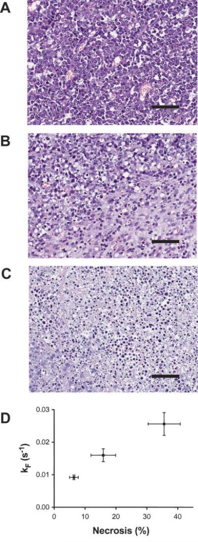
Nuclear spin hyperpolarization can dramatically increase the sensitivity of the (13)C magnetic resonance experiment, allowing dynamic measurements of the metabolism of hyperpolarized (13)C-labeled substrates in vivo. Here, we report a preclinical study of the response of lymphoma tumors to the vascular disrupting agent (VDA), combretastatin-A4-phosphate (CA4P), as detected by measuring changes in tumor metabolism of hyperpolarized [1-(13)C]pyruvate and [1,4-(13)C(2)]fumarate. These measurements were compared with dynamic contrast agent-enhanced magnetic resonance imaging (DCE-MRI) measurements of tumor vascular function and diffusion-weighted MRI (DW-MRI) measurements of the tumor cell necrosis that resulted from subsequent loss of tumor perfusion. The rate constant describing flux of hyperpolarized (13)C label between [1-(13)C]pyruvate and lactate was decreased by 34% within 6 hours of CA4P treatment and remained at this lower level at 24 hours. The rate constant describing production of labeled malate from hyperpolarized [1,4-(13)C(2)]fumarate increased 1.6-fold and 2.5-fold at 6 and 24 hours after treatment, respectively, and correlated with the degree of necrosis detected in histologic sections. Although DCE-MRI measurements showed a substantial reduction in perfusion at 6 hours after treatment, which had recovered by 24 hours, DW-MRI showed no change in the apparent diffusion coefficient of tumor water at 6 hours after treatment, although there was a 32% increase at 24 hours (P < 0.02) when regions of extensive necrosis were observed by histology. Measurements of hyperpolarized [1-(13)C]pyruvate and [1,4-(13)C(2)]fumarate metabolism may provide, therefore, a more sustained and sensitive indicator of response to a VDA than DCE-MRI or DW-MRI, respectively.
©2010 AACR.
Figures


References
-
- Tozer GM, Kanthou C, Baguley BC. Disrupting tumour blood vessels. Nat Rev Cancer. 2005;5:423–35. - PubMed
-
- Iyer S, Chaplin DJ, Rosenthal DS, Boulares AH, Li L, Smulson ME. Induction of Apoptosis in Proliferating Human Endothelial Cells by the Tumor-specific Antiangiogenesis Agent Combretastatin A-4. Cancer Res. 1998;58:4510–4. - PubMed
-
- Dark GG, Hill SA, Prise VE, Tozer GM, Pettit GR, Chaplin DJ. Combretastatin A-4, an agent that displays potent and selective toxicity toward tumor vasculature. Cancer Res. 1997;57:1829–34. - PubMed
-
- Galbraith SM, Maxwell RJ, Lodge MA, et al. Combretastatin A4 phosphate has tumor antivascular activity in rat and man as demonstrated by dynamic magnetic resonance imaging. J Clin Oncol. 2003;21:2831–42. - PubMed
Publication types
MeSH terms
Substances
Grants and funding
LinkOut - more resources
Full Text Sources
