. 2010 Dec 15;70(24):10161-9.
doi: 10.1158/0008-5472.CAN-10-1921.

The inflammasome component NLRP3 impairs antitumor vaccine by enhancing the accumulation of tumor-associated myeloid-derived suppressor cells

Affiliations

The inflammasome component NLRP3 impairs antitumor vaccine by enhancing the accumulation of tumor-associated myeloid-derived suppressor cells

Hendrik W van Deventer et al. Cancer Res. .

Abstract

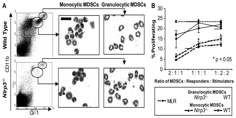
The inflammasome is a proteolysis complex that generates the active forms of the proinflammatory cytokines interleukin (IL)-1β and IL-18. Inflammasome activation is mediated by NLR proteins that respond to microbial and nonmicrobial stimuli. Among NLRs, NLRP3 senses the widest array of stimuli and enhances adaptive immunity. However, its role in antitumor immunity is unknown. Therefore, we evaluated the function of the NLRP3 inflammasome in the immune response using dendritic cell vaccination against the poorly immunogenic melanoma cell line B16-F10. Vaccination of Nlrp3(-/-) mice led to a relative 4-fold improvement in survival relative to control animals. Immunity depended on CD8(+) T cells and exhibited immune specificity and memory. Increased vaccine efficacy in Nlrp3(-/-) hosts did not reflect differences in dendritic cells but rather differences in myeloid-derived suppressor cells (MDSC). Although Nlrp3 was expressed in MDSCs, the absence of Nlrp3 did not alter either their functional capacity to inhibit T cells or their presence in peripheral lymphoid tissues. Instead, the absence of Nlrp3 caused a 5-fold reduction in the number of tumor-associated MDSCs found in host mice. Adoptive transfer experiments also showed that Nlrp3(-/-) MDSCs were less efficient in reaching the tumor site. Depleting MDSCs with an anti-Gr-1 antibody increased the survival of tumor-bearing wild-type mice but not Nlrp3(-/-) mice. We concluded that Nlrp3 was critical for accumulation of MDSCs in tumors and for inhibition of antitumor T-cell immunity after dendritic cell vaccination. Our findings establish an unexpected role for Nlrp3 in impeding antitumor immune responses, suggesting novel approaches to improve the response to antitumor vaccines by limiting Nlrp3 signaling.


Figures

Figure 1
Figure 1. Nlrp3 is expressed by tumor-associated myeloid cells
Expression of Nlrp3 by real time PCR in (1) Gr-1+, CD11b+ (2) Gr-1, CD11b+ and (3) Gr-1, CD11b sorted cells. The panel on left is a representative dot plot of sorted cells from a subcutaneous B16-F10 tumor in a vaccinated WT (A) and Nlrp3−/− mouse (B). The panel on the right is a bar graph of Nlrp3 transcripts normalized to cell number.
Figure 2
Figure 2. DC vaccine improves survival in Nlrp3−/− mice but not WT mice
(A) Survival curves of WT and Nlrp3−/− mice after receiving a subcutaneous injection with 1×10⁴ B16-F10 melanoma cells. (B) Survival of Nlrp3−/− mice was significantly improved after receiving 1×10⁶ tumor-lysate pulsed DCs (DC Vx) on day 3 and 10 after tumor injection. No improvement was seen in WT mice. (C) Nlrp3−/− mice show improved survival after receiving vaccines from either Nlrp3−/− or WT mice. (D) WT mice show no improvement in survival after receiving vaccines from WT or Nlrp3−/− mice.
Figure 3
Figure 3. Vaccinated Nlrp3−/− mice demonstrate greater CD8+ T cell response
(A) Survival curves of vaccinated (Vx) or naive Nlrp3−/− mice after rechallenge with B16-F10 or Lewis Lung cancer (LLCa) cells. (B) Survival of vaccinated Nlrp3−/− mice following depletion of CD8+, CD4+, and NK cells. (C) The graph shows the hazard ratio and confidence intervals that compare survival of cell depleted Nlrp3−/− with control Nlrp3−/− mice. Values greater than 1 (to the left) demonstrate decreased survival compared to untreated Nlrp3−/− mice. (D) Total number of CD4+ and CD8+ effector cells from DC vaccinated Nlrp3−/− (black) and WT (gray) mice. Palpable tumors (gray) were defined by a measurable difference (> 0.2 mm) between the right and left leg; non-palpable tumors were grossly identifiable during dissection.
Figure 4
Figure 4. WT and Nlrp3−/− MDSCs have a similar microscopic appearance and suppressive capacity
(A) Flow cytometry dot plots for tumor-associated cells isolated from vaccinated WT and Nlrp3−/− mice. Cells were sorted by the gates shown and then examined after cytospin with Wright Giemsa staining. Lens magnification is 500X; scale bar is 20 μm. (B) Line graph showing suppression of a MLR reaction by granulocytic (solid line) and monocytic (dashed line) MDSCs from WT (squares) and Nlrp3−/− (diamonds) mice. Significance was determined by comparing proliferation with control MLR response (dotted line). Results are averaged from three separate experiments.
Figure 5
Figure 5. Superior response to DC vaccines by Nlrp3−/− mice is due to impaired migration of MDSCs
(A) Percentages of MDSCs from the spleen and total number of MDSCs in the TDLN of WT (striped) and Nlrp3−/− (solid) mice as determined by flow cytometry 14 days after tumor injection. (B) The total number of tumor-associated Gr-1+, CD11b+ cells is increased in WT mice as determined by microscopy. (C) Increased percentage of monocytic MDSCs (left) and granulocytic MDSCs (right) from the tumor of WT (striped) compared to Nlrp3−/− (solid) mice as determined by flow cytometry. (D) Survival curves of WT and Nlrp3−/− mice treated with anti-Gr-1 antibody (dotted arrows) and DC vaccine (solid arrows). (E) Contour plots on the left show phenotype of MDSCs isolated from the spleens of WT (upper) and Nlrp3−/− (lower) mice prior to injection into Nlrp3−/− mice. Dot plots on the right show cells recovered from B16 tumors in vaccinated Nlrp3−/− mice. Upper panels are WT EGFP+ MDSCs and lower panels are Nlrp3−/− EGFP+ MDSCs. Values represent the average number of cells recovered from each tumor.

References

    1. Martinon F, Mayor A, Tschopp J. The Inflammasomes: Guardians of the body. Annu Rev Immunol. 2009;27:229–265. - PubMed
    1. Ting J, Lovering R, Alnemri E, et al. The NLR gene family: A standard nomenclature. Immunity. 2008;28:285–287. - PMC - PubMed
    1. Mariathasan S, Monack DM. Inflammasome adaptors and sensors: Intracellular regulators of infection and inflammation. Nat Rev Immunol. 2007;7:31–40. - PubMed
    1. Agostini L, Martinon F, Burns K, McDermott MF, Hawkins P, Tschopp J. NALP3 forms an IL-1beta-processing inflammasome with increased activity in Muckle-Wells autoinflammatory disorder. Immunity. 2004;20:319–25. - PubMed
    1. Kono H, Rock K. How dying cells alert the immune system to danger. Nat Rev Imm. 2008;8:279–289. - PMC - PubMed

Publication types

MeSH terms