. 2010 Dec 15;70(24):10340-50.
doi: 10.1158/0008-5472.CAN-10-0705.

Regulation of the embryonic morphogen Nodal by Notch4 facilitates manifestation of the aggressive melanoma phenotype

Affiliations

Regulation of the embryonic morphogen Nodal by Notch4 facilitates manifestation of the aggressive melanoma phenotype

Katharine M Hardy et al. Cancer Res. .

Abstract

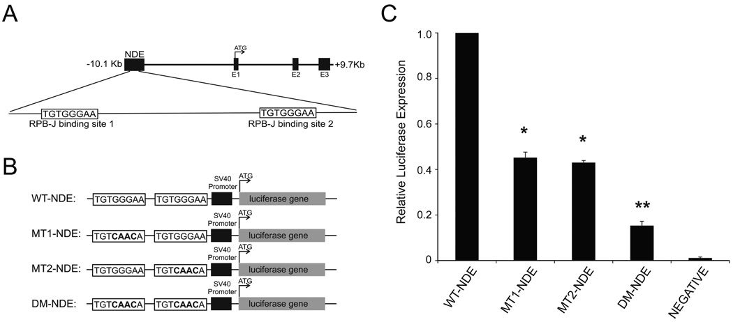
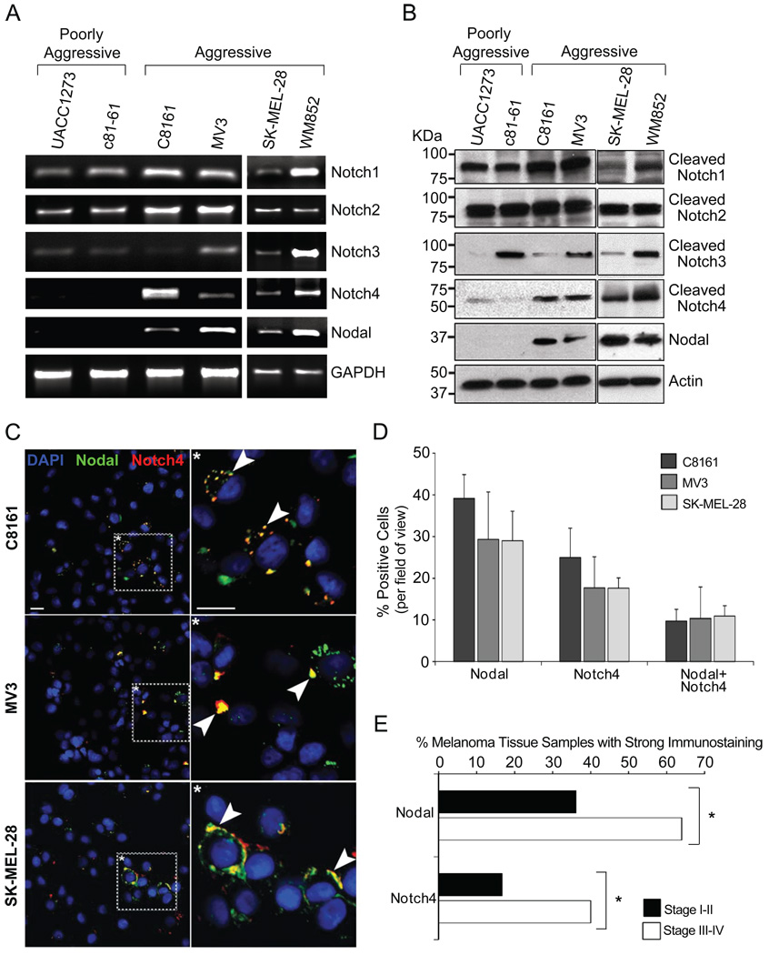
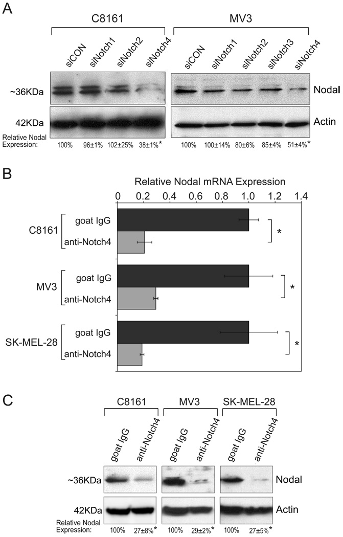
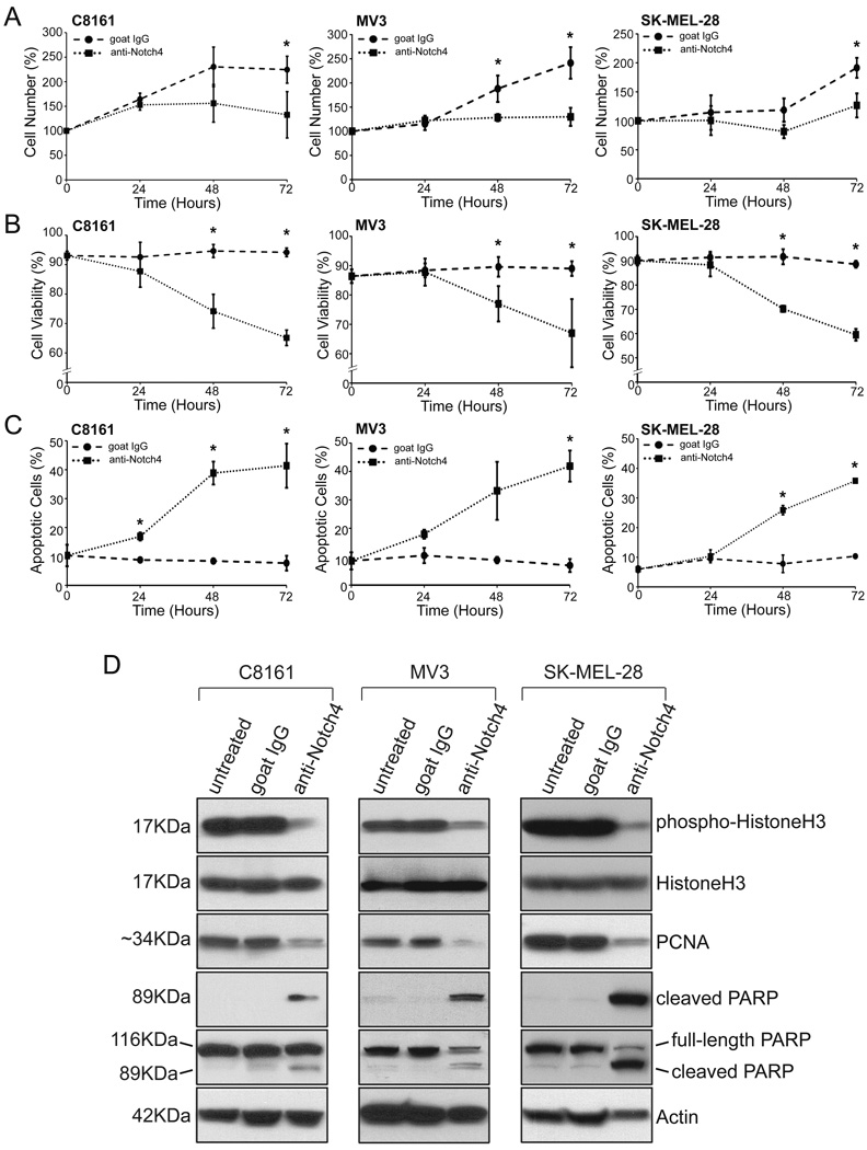
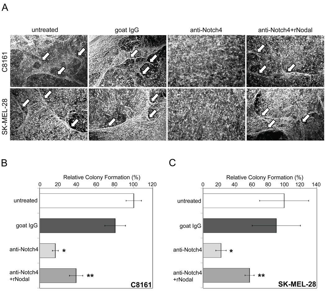
Metastatic melanoma is an aggressive skin cancer associated with poor prognosis. The reactivation of the embryonic morphogen Nodal in metastatic melanoma has previously been shown to regulate the aggressive behavior of these tumor cells. During the establishment of left-right asymmetry in early vertebrate development, Nodal expression is specifically regulated by a Notch signaling pathway. We hypothesize that a similar relationship between Notch and Nodal may be reestablished in melanoma. In this study, we investigate whether cross talk between the Notch and Nodal pathways can explain the reactivation of Nodal in aggressive metastatic melanoma cells. We show a molecular link between Notch and Nodal signaling in the aggressive melanoma cell line MV3 via the activity of an RBPJ-dependent Nodal enhancer element. We show a precise correlation between Notch4 and Nodal expression in multiple aggressive cell lines but not poorly aggressive cell lines. Surprisingly, Notch4 is specifically required for expression of Nodal in aggressive cells and plays a vital role both in the balance of cell growth and in the regulation of the aggressive phenotype. In addition, Notch4 function in vasculogenic mimicry and anchorage-independent growth in vitro is due in part to Notch4 regulation of Nodal. This study identifies an important role for cross talk between Notch4 and Nodal in metastatic melanoma, placing Notch4 upstream of Nodal, and offers a potential molecular target for melanoma therapy.


Conflict of interest statement

Potential conflicts of interest: Commercial research grant from Acceleron Pharma (M.J.C. Hendrix); Patent to target Nodal (M.J.C. Hendrix, E.A. Seftor, L.M. Postovit)

Figures

Figure 1
Figure 1
A Nodal enhancer element drives luciferase expression in MV3 cells in an RBPJ-dependent manner. A) Representation of the Nodal gene locus with upstream Node-specific enhancer (NDE) region and the two putative RBP-J binding site. B) Wild-type (WT-NDE), single mutant (MT1-NDE; MT2-NDE) and double mutant (DM-NDE) reporter constructs utilized. C) The ability of wild-type and mutant NDE constructs to drive luciferase activity in MV3 cells. pGL3-basic (NEGATIVE) was a negative control.*Significant difference from WT-NDE (p<0.05);**significant difference from MT1-NDE/MT2-NDE (p<0.05). Graph is an average of three independent experiments (+SEM).
Figure 2
Figure 2
Survey of Notch receptors and Nodal expression in melanoma cell lines. A) RNA isolated from two poorly aggressive cell lines (UACC1273, c81-61) and four aggressive cell lines (C8161, MV3, SK-MEL-28, WM852) was assayed for gene expression of Notch1–4 and Nodal by semi-quantitative PCR. GAPDH was a loading control. DNA contamination was excluded using no MMLV (not shown). B) Protein lysates were analyzed for Notch receptor and Nodal proteins by Western blotting. Actin was a loading control. C) Nodal (green) and Notch4 (red) proteins were examined by confocal microscopy in C8161, MV3, and SK-MEL-28 cells (also Supplemental Fig.1). Arrowheads in inset (*) denote regions of co-localization (yellow). DAPI (blue) marks cell nuclei. White bar represents 10µm. D) Cells positive for Nodal, Notch4, or both Nodal and Notch4 (Nodal+Notch4) were independently counted using a 25X objective. For each category, mean+/−SD was graphed as a percentage of total DAPI-positive nuclei (n=7). E) Immunohistochemistry for Nodal and Notch4 was performed on serial sections of a human melanoma tissue array. The number of tissue samples showing strong staining (>50%) was graphed as a percentage of total samples evaluated (for Stage I–II, n=36; for Stage III–IV, n=25).*p<0.05.
Figure 3
Figure 3
Notch4 signaling regulates expression of Nodal. A) Transfection of C8161 (left) and MV3(right) cells with siRNA to Notch1 (siNotch1), Notch2 (siNotch2), Notch3 (siNotch3), Notch4 (siNotch4), or a negative control (siCON), followed by Western blotting for Nodal protein. Actin was a loading control. Relative Nodal expression was determined by densitometry (n=3). B–C) C8161, MV3, and SK-MEL-28 cells were treated with anti-human Notch4 neutralizing antibody or IgG. RNA was analyzed by real-time PCR for expression of Nodal mRNA (B). Protein lysates were analyzed for Nodal and Actin (C), and relative Nodal expression evaluated by densitometry (n=3).*p<0.05.
Figure 4
Figure 4
Inhibition of Notch4 activity limits cell proliferation and promotes apoptosis. C8161, MV3, and SK-MEL-28 cells were treated with anti-human Notch4 antibody or IgG. A–C) At 24-hour time points, cells were assayed for cell number (A), viability (B), and apoptosis (C) by flow cytometry. Cell number is shown as a percentage of the initial population, while viability and apoptosis is represented as a percentage of total cells. Plots represent the mean of three independent experiments performed in triplicate (+/−SD);*p<0.05. D) Western blot analyses of HistoneH3 phosphorylation and PCNA expression (proliferation), and PARP cleavage (apoptosis) in C8161, MV3, and SK-MEL-28 cells. Actin was a loading control. Membranes were stripped between antibody detections. Western blots are representative of three experiments. Abbreviations: PCNA, Proliferating Cell Nuclear Antigen; PARP, Poly ADP-Ribosome Polymerase.
Figure 5
Figure 5
Inhibition of Notch4 signaling blocks vasculogenic mimicry and anchorage independent growth in vitro in a Nodal-dependent manner. A) C8161 and SK-MEL-28 cells were seeded on 3D-collagen gel matrix and left untreated or treated with IgG, anti-Notch4 antibody, or anti-Notch4 antibody plus recombinant human Nodal (rNodal). White arrows indicate vascular-like network formation in untreated (far-left), IgG-treated (center-left), and anti-Notch4+rNodal-treated cultures (far-right). Original magnification 100X. B–C) Relative colony formation in C8161 (B) and SK-MEL-28 (C) cells cultured on soft agar following pretreatment with IgG or anti-Notch4 antibody with or without rNodal. Graph indicates macroscopic colonies as a percentage of control (+/− SD).*Significant difference from untreated/IgG-treated cultures (p<0.05);**significant difference from anti-Notch4 treated cultures (p<0.05). Graphs depict one representative experiment of three.

References

    1. Hendrix MJ, Seftor EA, Seftor RE, Kasemeier-Kulesa J, Kulesa PM, Postovit LM. Reprogramming metastatic tumour cells with embryonic microenvironments. Nat Rev Cancer. 2007;7:246–255. - PubMed
    1. Postovit LM, Margaryan NV, Seftor EA, et al. Human embryonic stem cell microenvironment suppresses the tumorigenic phenotype of aggressive cancer cells. Proc Natl Acad Sci U S A. 2008;105:4329–4334. - PMC - PubMed
    1. Topczewska JM, Postovit LM, Margaryan NV, et al. Embryonic and tumorigenic pathways converge via Nodal signaling: role in melanoma aggressiveness. Nat Med. 2006;12:925–932. - PubMed
    1. Massi D, Tarantini F, Franchi A, et al. Evidence for differential expression of Notch receptors and their ligands in melanocytic nevi and cutaneous malignant melanoma. Mod Pathol. 2006;19:246–254. - PubMed
    1. Pinnix CC, Lee JT, Liu ZJ, et al. Active Notch1 confers a transformed phenotype to primary human melanocytes. Cancer Res. 2009;69:5312–5320. - PMC - PubMed

Publication types

MeSH terms