. 2011 Feb 18;286(7):5607-13.
doi: 10.1074/jbc.M110.188433. Epub 2010 Dec 15.

Structural basis for three-step sequential catalysis by the cholesterol side chain cleavage enzyme CYP11A1

Affiliations

Structural basis for three-step sequential catalysis by the cholesterol side chain cleavage enzyme CYP11A1

Natalia Mast et al. J Biol Chem. .

Abstract

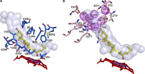
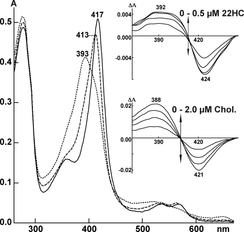
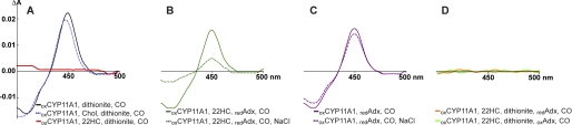
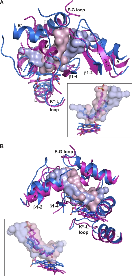
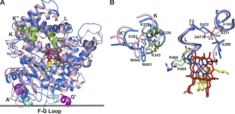
Mitochondrial cytochrome P450 11A1 (CYP11A1 or P450 11A1) is the only known enzyme that cleaves the side chain of cholesterol, yielding pregnenolone, the precursor of all steroid hormones. Pregnenolone is formed via three sequential monooxygenation reactions that involve the progressive production of 22R-hydroxycholesterol (22HC) and 20α,22R-dihydroxycholesterol, followed by the cleavage of the C20-C22 bond. Herein, we present the 2.5-Å crystal structure of CYP11A1 in complex with the first reaction intermediate, 22HC. The active site cavity in CYP11A1 represents a long curved tube that extends from the protein surface to the heme group, the site of catalysis. 22HC occupies two-thirds of the cavity with the 22R-hydroxyl group nearest the heme, 2.56 Å from the iron. The space at the entrance to the active site is not taken up by 22HC but filled with ordered water molecules. The network formed by these water molecules allows the "soft" recognition of the 22HC 3β-hydroxyl. Such a mode of 22HC binding suggests shuttling of the sterol intermediates between the active site entrance and the heme group during the three-step reaction. Translational freedom of 22HC and torsional motion of its aliphatic tail are supported by solution studies. The CYP11A1-22HC co-complex also provides insight into the structural basis of the strict substrate specificity and high catalytic efficiency of the enzyme and highlights conserved structural motifs involved in redox partner interactions by mitochondrial P450s.


Figures

FIGURE 1.
FIGURE 1.
Views of the CYP11A1 active site illustrating interactions with 22HC. A, amino acid residues (marine) within 4 Å of 22HC. B, array of structural water molecules (violet spheres) hydrogen-bonded to the 22HC 3β-hydroxyl and to the residues (pink) at the entrance to the active site. For clarity, residues interacting with 22HC are omitted in this panel. The heme group is depicted in red, and 22HC is depicted in yellow. The nitrogen, oxygen, sulfur, and iron atoms are in blue, red, yellow, and orange, respectively. The enclosed volume of the active site is shown as a semitransparent surface.
FIGURE 2.
FIGURE 2.
Absolute and difference (insets) spectra of CYP11A1 in the absence (solid line) and presence of 22HC (dashed line) and cholesterol (dotted line). Absolute spectra were recorded using 5 μm CYP11A1 with the concentrations of 22HC and cholesterol being 50 μm. Both sterols were added from 10 mm stocks in 45% aqueous 2-hydroxypropyl-β-cyclodextrin. The buffer was 50 mm potassium phosphate (pH 7.2) containing 1 mm EDTA. Difference spectra in the insets were recorded using 0.4 μm CYP11A1. 22HC was added from 0.1 mm stock in 4.5% aqueous 2-hydroxypropyl-β-cyclodextrin, and cholesterol (Chol.) was added from 0.5 mm stock 4.5% aqueous 2-hydroxypropyl-β-cyclodextrin.
FIGURE 3.
FIGURE 3.
Binding of CO to reduced CYP11A1 as assessed by difference spectroscopy (Fe2+-CO versus Fe2+-P450 spectrum). By utilizing different assay conditions, we show that blockage of CO binding by 22HC is relieved by Adx binding, and that upon release of Adx, 22HC is able to significantly displace bound CO. The same concentration of P450 (0.25 μm) was used in each experiment, and the order in which reagents were mixed is indicated; the ox and red subscripts specify whether CYP11A1 and Adx are in oxidized and reduced states, respectively. A, reduction of CYP11A1 with sodium dithionite in the absence (black line) and presence (red line) of equimolar 22HC and a 10-fold molar excess of cholesterol (Chol; dashed blue line). B, reduction of CYP11A1 with reduced Adx in the presence of equimolar 22HC (solid green line) and in the presence of equimolar 22HC followed by addition of 1 m NaCl (dashed green line). C, control experiment in which CYP11A1 was treated either with reduced Adx only (solid magenta line) or with reduced Adx followed by addition of 1 m NaCl (dashed magenta line). D, control experiment in which the CYP11A1–22HC complex was reduced with sodium dithionite, followed by addition of reduced (orange line) or oxidized (green line) Adx.
FIGURE 4.
FIGURE 4.
Superimposed views of the active sites in CYP11A1 and CYP46A1 illustrating positioning of the secondary structural elements (A and B) and sterol substrates (insets). Secondary structural elements and the heme group are colored in marine in CYP11A1 and magenta in CYP46A1. The solvent-occupied volume of the active site is shown either as a solid or semitransparent surface in light blue in CYP11A1 and light pink in CYP46A1. 22HC is in blue, and cholesterol 3-sulfate is in magenta. Coloring of atoms is the same as in Fig. 1.
FIGURE 5.
FIGURE 5.
Superpositioning of CYP11A1 (marine) and CYP24A1 (light pink) illustrating interactions with the membrane and Adx. A, comparison of the overall fold at the proximal side showing the positions of the membrane insertion sequences (cyan in CYP11A1 and magenta in CYP24A1) and of the secondary structural elements that could participate in Adx binding in mitochondrial P450s (K, K″, and L helices, in green). The gray dotted line separates the cytosol (above) and the lipid bilayer (below) with respect to CYP11A1. B, conserved positively charged residues (dark and light green in CYP11A1 and CYP24A1, respectively) and tertiary structure interactions that govern specificity for the redox partner in mitochondrial P450s. One such interaction involves the invariant glutamic acid in the K helix (Glu-383), which hydrogen bonds with the invariant tryptophan in the K″ helix (Trp-440) in CYP24A1 (10); in CYP11A1, this interaction exists as the Glu-344–Trp-401 hydrogen bond. In addition, Glu-432 in CYP11A1 and its counterpart Gln-471 in CYP24A1, at the N terminus of the L helix, are hydrogen-bonded to the spatially overlaid structural Wat-532 and Wat-516, respectively, which in turn interact with residues in the I (Gly-288) and E (Ala-168) helices in CYP11A1 and with Ser-211 in the E helix in CYP24A1. Coloring of atoms is the same as in Fig. 1.

References

    1. Simpson E. R., Boyd G. S. (1967) Eur. J. Biochem. 2, 275–285 - PubMed
    1. Burstein S., Middleditch B. S., Gut M. (1975) J. Biol. Chem. 250, 9028–9037 - PubMed
    1. Orme-Johnson N. R., Light D. R., White-Stevens R. W., Orme-Johnson W. H. (1979) J. Biol. Chem. 254, 2103–2111 - PubMed
    1. Lambeth J. D., Kitchen S. E., Farooqui A. A., Tuckey R., Kamin H. (1982) J. Biol. Chem. 257, 1876–1884 - PubMed
    1. Pikuleva I. A. (2006) Drug Metab. Dispos. 34, 513–520 - PubMed

Publication types

MeSH terms

Substances

Associated data

LinkOut - more resources