. 2011 Mar 1;20(5):975-87.
doi: 10.1093/hmg/ddq543. Epub 2010 Dec 15.

TOPORS, implicated in retinal degeneration, is a cilia-centrosomal protein

Affiliations

TOPORS, implicated in retinal degeneration, is a cilia-centrosomal protein

Christina F Chakarova et al. Hum Mol Genet. .

Abstract

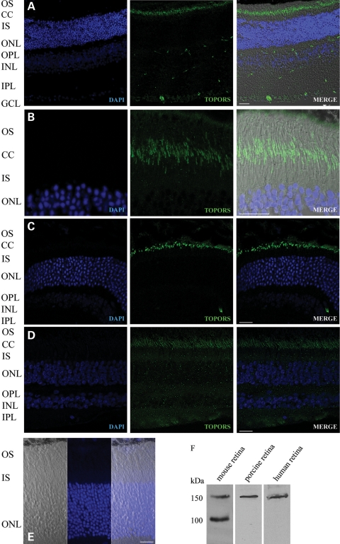
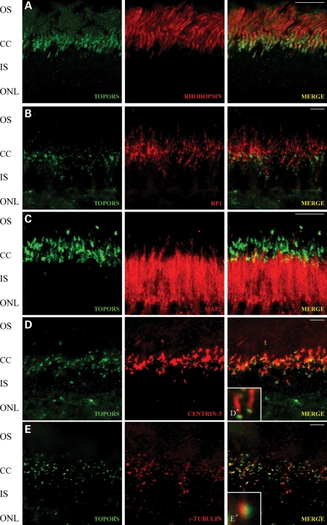
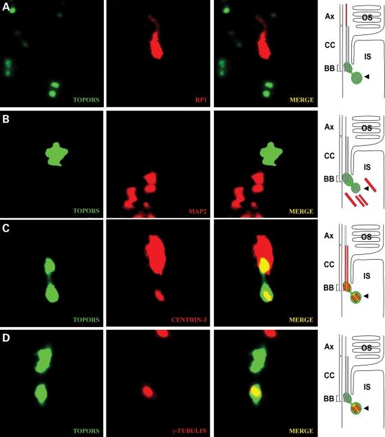
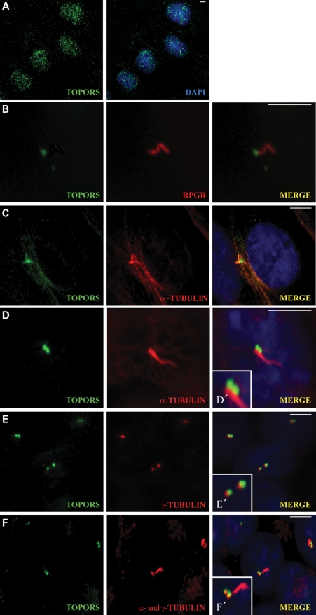
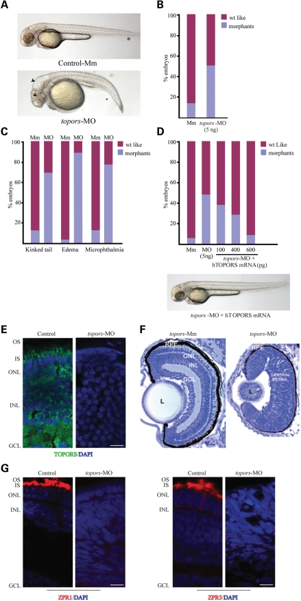
We recently reported that mutations in the widely expressed nuclear protein TOPORS (topoisomerase I-binding arginine/serine rich) are associated with autosomal dominant retinal degeneration. However, the precise localization and a functional role of TOPORS in the retina remain unknown. Here, we demonstrate that TOPORS is a novel component of the photoreceptor sensory cilium, which is a modified primary cilium involved with polarized trafficking of proteins. In photoreceptors, TOPORS localizes primarily to the basal bodies of connecting cilium and in the centrosomes of cultured cells. Morpholino-mediated silencing of topors in zebrafish embryos demonstrates in another species a comparable retinal problem as seen in humans, resulting in defective retinal development and failure to form outer segments. These defects can be rescued by mRNA encoding human TOPORS. Taken together, our data suggest that TOPORS may play a key role in regulating primary cilia-dependent photoreceptor development and function. Additionally, it is well known that mutations in other ciliary proteins cause retinal degeneration, which may explain why mutations in TOPORS result in the same phenotype.


Figures

Figure 1.
Figure 1.
Localization of TOPORS in the retina. (A–D) Indirect immunofluorescence analysis of mouse, porcine and human retina sections using anti-TOPORS antibody (green). (A) TOPORS is localized only in the ciliary region of the photoreceptors and in the nuclei of the ganglion cells in the mouse retina. (B) Higher magnification only of the photoreceptor cell layer of the mouse retina. (C) Immunostaining of anti-TOPORS antibody in the porcine retina. (D) Immunostaining of anti-TOPORS antibody in the human retina. (E) As a negative control, the mouse section was probed with TOPORS antibody blocked with TOPORS recombinant protein (206 amino acid), where no signal could be observed. (F) Immunoblot analysis of the specificity of mouse anti-TOPORS antibody shows a 150 kDa signal detected in mouse, porcine and human retina lysates, where, in the mouse, two bands could be seen with the molecular weight of 100 and 150 kDa due to a specific smaller isoform in this species lacking exon 2 of the gene. OS, outer segment; CC, connecting cilium; IS, inner segment; ONL, outer nuclear layer; INL, inner nuclear layer; GCL, ganglion cell layer. Nuclei are stained with DAPI (blue). Scale bar: 10 μm.
Figure 2.
Figure 2.
Subcellular localization of TOPORS at the ciliary region of the photoreceptor cell. (AE) Indirect immunofluorescence analysis of mouse retina sections stained with anti-TOPORS antibody (green) with ciliary proteins and proteins of the outer and inner photoreceptor segments (red) in the retina. (A) Photoreceptor localization of TOPORS with the most abundant protein in these cells in the area of the outer segment—rhodopsin. (B) Subcellular localization of TOPORS and RP1 as a marker for photoreceptor ciliary axoneme. TOPORS is localized at the region of the connecting cilium (CC) of the photoreceptor cells. (C) Double-labeling with anti-TOPORS and anti-MAP2. Antibodies against TOPORS stain the CC region. MAP2 is present in the inner segment (IS) of the photoreceptor cell and slightly at the CC region. (D) Indirect immunofluorescence double-labeling with antibodies against TOPORS and the marker of the CC, basal body (BB) and centriole centrin (Centrin-3) in cryosections through mouse retinas. (D′) Higher magnification of merged signal (TOPORS and Centrin-3) from the CC of a single photoreceptor cell. (E) Double-staining with anti-TOPORS and anti-γ-tubulin. TOPORS is localized at the cilium of the photoreceptor cell. Anti-γ-tubulin stains the basal body. (E′) Higher magnification of merged signal of TOPORS and γ-tubulin, which partly co-localizes in the basal body of the CC of a single photoreceptor cell. Scale bar: 10 μm. OS, outer segment; CC, connecting cilia; IS, inner segment; ONL, outer nuclear layer; INL, inner nuclear layer; GCL, ganglion cell layer.
Figure 3.
Figure 3.
TOPORS is localized at the basal body and centriole of the photoreceptor cell. (AD) Indirect immunofluorescence double-staining with antibodies against TOPORS and marker proteins for the ciliary region of cryosections in mouse retinas. High-magnification images of double-immunofluorescences of the ciliary region of photoreceptor cells and schematic presentations (on the left) illustrating subciliary localization of TOPORS (green) and marker molecules (red). (A) Anti-TOPORS and anti-RP1 immunolabeling. RP1 is used as a marker indicating the ciliary axoneme (Ax). TOPORS and RP1 do not co-localize. (B) Anti-TOPORS and anti-MAP2 immunolabeling. MAP2 stains the microtubules in the inner segment (IS) of the photoreceptor cells. (C) Centrin-3 labels connecting cilium (CC), basal body (BB) and centriole (arrowhead). According to this centrin-3 labeling, TOPORS is present at the BB and at the centriole (arrowhead). (D) Anti-TOPORS and anti-γ-tubulin immunostaining. γ-Tubulin stains the centriole (arrowhead). TOPORS partly co-localize with γ-tubulin at the BB. OS, outer segment of the photoreceptor cell; IS, inner segment of the photoreceptor cell.
Figure 4.
Figure 4.
TOPORS localizes to the basal body of the primary cilia and associates with microtubule proteins in ciliated cells. (A) Endogenous TOPORS (green) localized in the nucleus stained with DAPI (blue) of mitotic MDCK cells. (B) TOPORS (green) localizes to the base of the ciliary axoneme stained with RPGR (red) in ciliated MDCK cells. (C) In ciliated ARPE-19 cells, TOPORS (green) localizes to the base of the cilium; the ciliary axoneme is stained with α-tubulin (red). (D) In ciliated IMCD3 cells, TOPORS (green) localizes to the base of the cilium; the ciliary axoneme is stained with α-tubulin (red). (D′) Higher magnification of the ciliary structure. (E) Double-staining of ciliated IMCD3 cells with TOPORS (green) and γ-tubulin (red) used as a marker for the basal bodies. Both proteins localize in the same area with some overlap but do not co-localize entirely (see also Supplementary Material, Fig. S3). (E′) Higher magnification of the basal body. (F) Ciliated IMCD3 cells stained with TOPORS (green), a- and γ-tubulin (both in red), highlighting the basal bodies and ciliary axoneme. (F′) Higher magnification of the ciliary apparatus. Nuclei are stained with DAPI. Scale bar: 10 μm.
Figure 5.
Figure 5.
Localization of TOPORS during cell cycle. (A) RPE-1 cells were transfected with plasmid encoding for TOPORS-GFP and GFP alone, fixed and stained for the centrosomal marker γ-tubulin. (B and C) Synchronized RPE-1 cells were fixed and stained for endogenous TOPORS and γ-tubulin. G0 was stained using anti-acetylated α-tubulin antibody. DNA was stained with DAPI. Insets show higher magnifications of centrosome-containing regions. Representative images of each step of the cell cycle are shown. In colored images, TOPORS staining is in green, centrosomes in red, nuclei in blue and the arrow shows the mid-body. Scale bar: 10 μm.
Figure 6.
Figure 6.
Knockdown of zebrafish topors causes developmental anomalies and the effect of the inhibition of topors on zebrafish retina. (A) Injection of anti-sense morpholino (topors-MO) into zebrafish embryos results in developmental disorders, including shortened body axis and hydrocephaly. Embryos injected with the 5 base mismatch (Mm) control are also shown. Asterisk depicts kinked tail; long arrow shows microphthalmia; arrowhead indicates hydrocephaly. (B and C) Histograms showing the frequency of the occurrence of morphants. (B) Incidence of the different phenotypes observed in the MO-treated embryos compared with Mm-treated controls. (C) Results are representative of at least three independent experiments. (D) The topors-knockdown phenotype can be rescued by injecting indicated doses of human TOPORS (hTOPORS) mRNA. Data are representative of three independent experiments (n > 100). Bottom panel shows representative image of an embryo co-injected with the specific MO against topors and mRNA encoding hTOPORS. Majority of the phenotypes seem to be rescued by human TOPORS. (E) Immunohistochemistry of cryosections of 4 dpf zebrafish retina was performed using anti-TOPORS antibody (green). DAPI was used to stain the nuclei (blue). IS, inner segment; OS, outer segment; ONL, outer nuclear layer; INL, inner nuclear layer; GCL, ganglion cell layer. (F) Retinal histology of control (Mm) and topors-MO injected (MO) embryos at 4 dpf (×40). Plastic sections from 4 dpf zebrafish embryo were stained with toludine blue. (G) Cryosections of 4 dpf zebrafish were immunostained with Zpr1 or Zpr3 antibody (red) for marking cone and rod photoreceptors, respectively. Nuclear layers are stained with DAPI (blue). Scale bar: 10 μm.

References

    1. Hartong D.T., Berson E.L., Dryja T.P. Retinitis pigmentosa. Lancet. 2006;368:1795–1809. doi:10.1016/S0140-6736(06)69740-7. - DOI - PubMed
    1. Wright A.F., Chakarova C.F., Abd El-Aziz M.M., Bhattacharya S.S. Photoreceptor degeneration: genetic and mechanistic dissection of a complex trait. Nat. Rev. Genet. 2010;11:273–284. doi:10.1038/nrg2717. - DOI - PubMed
    1. Rivolta C., Sharon D., DeAngelis M.M., Dryja T.P. Retinitis pigmentosa and allied diseases: numerous diseases, genes, and inheritance patterns. Hum. Mol. Genet. 2002;11:1219–1227. doi:10.1093/hmg/11.10.1219. - DOI - PubMed
    1. Chakarova C.F., Papaioannou M.G., Khanna H., Lopez I., Waseem N., Shah A., Theis T., Friedman J., Maubaret C., Bujakowska K., et al. Mutations in TOPORS cause autosomal dominant retinitis pigmentosa with perivascular RPE atrophy. Am. J. Hum. Genet. 2007;81:1098–1103. doi:10.1086/521953. - DOI - PMC - PubMed
    1. Bowne S.J., Sullivan L.S., Gire A.I., Birch D.G., Hughbanks-Wheaton D., Heckenlively J.R., Daiger S.P. Mutations in the TOPORS gene cause 1% of autosomal dominant retinitis pigmentosa. Mol. Vis. 2008;14:922–927. - PMC - PubMed

Publication types

MeSH terms