Evaluation of dose to cardiac structures during breast irradiation
- PMID: 21159806
- PMCID: PMC3473433
- DOI: 10.1259/bjr/12497075
Evaluation of dose to cardiac structures during breast irradiation
Abstract
Objective: Adjuvant radiotherapy for breast cancer can lead to late cardiac complications. The highest radiation doses are likely to be to the anterior portion of the heart, including the left anterior descending coronary artery (LAD). The purpose of this work was to assess the radiation doses delivered to the heart and the LAD in respiration-adapted radiotherapy of patients with left-sided breast cancer.
Methods: 24 patients referred for adjuvant radiotherapy after breast-conserving surgery for left-sided lymph node positive breast cancer were evaluated. The whole heart, the arch of the LAD and the whole LAD were contoured. The radiation doses to all three cardiac structures were evaluated.
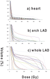
Results: For 13 patients, the plans were acceptable based on the criteria set for all 3 contours. For seven patients, the volume of heart irradiated was well below the set clinical threshold whereas a high dose was still being delivered to the LAD. In 1 case, the dose to the LAD was low while 19% of the contoured heart volume received over 20 Gy. In five patients, the dose to the arch LAD was relatively low while the dose to the whole LAD was considerably higher.
Conclusion: This study indicates that it is necessary to assess the dose delivered to the whole heart as well as to the whole LAD when investigating the acceptability of a breast irradiation treatment. Assessing the dose to only one of these structures could lead to excessive heart irradiation and thereby increased risk of cardiac complications for breast cancer radiotherapy patients.
Figures


References
-
- Clarke M, Collins R, Darby S, Davies C, Elphinstone P, Evans E, et al. Effects of radiotherapy and of differences in the extent of surgery for early breast cancer on local recurrence and 15-year survival: an overview of the randomised trials. Lancet 2005;366:2087–106 - PubMed
-
- Lind PARM, Wennberg B, Gagliardi G, Fornander T. Pulmonary complications following different radiotherapy techniques for breast cancer, and the association to irradiated lung volume and dose. Breast Cancer Res Treat 2001;68:199–210 - PubMed
-
- Correa CR, Das IJ, Litt HI, Ferrari V, Hwang WT, Solin LJ, et al. Association between tangential beam treatment parameters and cardiac abnormalities after definitive radiation treatment for left-sided breast cancer. Int J Radiat Oncol Biol Phys 2008;72:508–16 - PubMed
-
- Harris EE. Cardiac mortality and morbidity after breast cancer treatment. Cancer Control 2008;15:120–9 - PubMed
-
- Taylor CW, Nisbet A, McGale P, Goldman U, Darby SC, Hall P, et al. Cardiac doses from Swedish breast cancer radiotherapy since the 1950s. Radiother Oncol 2009;90:127–35 - PubMed
MeSH terms
LinkOut - more resources
Full Text Sources
Medical

