. 2011 Mar;85(5):2201-11.
doi: 10.1128/JVI.01748-10. Epub 2010 Dec 15.

Serotype-specific inactivation of the cellular DNA damage response during adenovirus infection

Affiliations

Serotype-specific inactivation of the cellular DNA damage response during adenovirus infection

Natalie A Forrester et al. J Virol. 2011 Mar.

Abstract

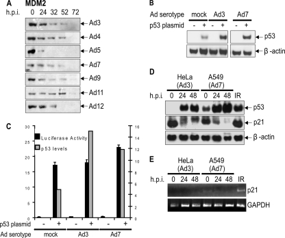
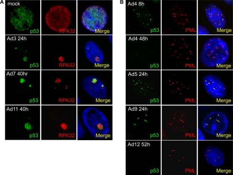
Adenovirus type 5 (Ad5) inactivates the host cell DNA damage response by facilitating the degradation of Mre11, DNA ligase IV, and p53. In the case of p53, this is achieved through polyubiquitylation by Ad5E1B55K and Ad5E4orf6, which recruit a Cul5-based E3 ubiquitin ligase. Recent evidence indicates that this paradigm does not apply to other adenovirus serotypes, since Ad12, but not Ad5, causes the degradation of TOPBP1 through the action of E4orf6 alone and a Cul2-based E3 ubiquitin ligase. We now have extended these studies to adenovirus groups A to E. While infection by Ad4, Ad5, and Ad12 (groups E, C, and A, respectively) cause the degradation of Mre11, DNA ligase IV, and p53, infection with Ad3, Ad7, Ad9, and Ad11 (groups B1, B1, D, and B2, respectively) only affects DNA ligase IV levels. Indeed, Ad3, Ad7, and Ad11 cause the marked accumulation of p53. Despite this, MDM2 levels were very low following infection with all of the viruses examined here, regardless of whether they increase p53 expression. In addition, we found that only Ad12 causes the degradation of TOPBP1, and, like Ad5, Ad4 recruits a Cul5-based E3 ubiquitin ligase to degrade p53. Surprisingly, Mre11 and DNA ligase IV degradation do not appear to be significantly affected in Ad4-, Ad5-, or Ad12-infected cells depleted of Cul2 or Cul5, indicating that E1B55K and E4orf6 recruit multiple ubiquitin ligases to target cellular proteins. Finally, although Mre11 is not degraded by Ad3, Ad7, Ad9, and Ad11, no viral DNA concatemers could be detected. We suggest that group B and D adenoviruses have evolved mechanisms based on the loss of DNA ligase IV and perhaps other unknown molecules to disable the host cell DNA damage response to promote viral replication.


Figures

FIG. 1.
FIG. 1.
Expression of Mre11, DNA ligase IV, p53, and TOPBP1 following viral infection. HeLa cells were infected with Ad3, Ad4, Ad5, Ad7, Ad9, Ad11, and Ad12. Cells were harvested at the times shown and subjected to Western blotting for Mre11 (A), DNA ligase IV (B), p53 (C), and TOPBP1 (D).
FIG. 2.
FIG. 2.
Effect of ablation of Cul2 and Cul5 expression on protein degradation during viral infection. HeLa cells were treated with nonsilencing siRNAs (non-sil) or siRNAs against Cul2 or Cul5. Cells then were infected with Ad4 (A), Ad5 (B), and Ad12 (C). Cells were harvested at the times shown and Western blotted with the indicated antibodies.
FIG. 3.
FIG. 3.
Transcriptional activity of p53 after viral infection. (A) HeLa cells were infected with Ad3, Ad4, Ad5, Ad7, Ad9, Ad11, and Ad12, harvested at the times shown, and Western blotted for MDM2. H1299 cells were transfected with a luciferase reporter construct (see Materials and Methods) and cDNA expressing p53. They then were infected with Ad3 or Ad7 and harvested after 24 h, and cells were Western blotted for p53 expression (B) and luciferase activity was determined (C). Relative p53 levels are shown in grey columns, and relative luciferase activity is shown in black columns (D and E). HeLa and A549 cells were infected with Ad3 and Ad7 as indicated. Samples were Western blotted for p53, p21, and β-actin (D) and subjected to RT-PCR for p21 (E). Glyceraldehyde-3-phosphate dehydrogenase (GAPDH) is included as a loading control. The right lane in D and E shows A549 cells treated with ionizing radiation (3 Gy) after 2 h.
FIG. 4.
FIG. 4.
Localization of Mre11 following adenovirus infection. HeLa cells were infected with Ad5 and Ad9 (A), Ad3, Ad7, Ad11, and Ad12 (B), and Ad4 (C) for the times shown. Cells were stained for Mre11 (green), PML (red), and RPA32 (red) as indicated and viewed by confocal microscopy as described in the text. Merged images are shown in the right column of images together with DAPI-stained DNA.
FIG. 5.
FIG. 5.
Localization of p53 following adenovirus infection. HeLa cells were infected with Ad3, Ad7, and Ad11 (A) and Ad4, Ad5, Ad9, and Ad12 (B) for the times shown. Cells were stained for p53 (green), RPA32 (red), or PML (red) and viewed by confocal microscopy as described in the text. Merged images are shown in the right column of images together with DAPI-stained DNA.
FIG. 6.
FIG. 6.
Localization of TOPBP1 following adenovirus infection. HeLa cells were infected with Ad3, Ad4, Ad5, and Ad7 (A) and Ad9, Ad11, and Ad12 (B) for the times shown. Cells were stained for RPA32 (green) and TOPBP1 (red) and viewed by confocal microscopy as described in the text. Merged images are shown in the right column together with DAPI-stained DNA.
FIG. 7.
FIG. 7.
Phosphorylation of Chk1 following adenovirus infection. HeLa cells were infected with Ad3, Ad4, Ad5, Ad7, Ad9, Ad11, and Ad12, harvested at the times shown, and Western blotted for phospho-Chk1 (Ser345) (A) and Chk1 (B). The final track in the Ad12 phospho-Chk1 blots shows HeLa cell extracts treated with hydroxyurea (HU) as a positive control.
FIG. 8.
FIG. 8.
Phosphorylation of KAP1 during adenoviral infection. HeLa cells were infected with Ad3, Ad4, Ad5, Ad7, Ad9, Ad11, and Ad12, harvested at the times shown, and Western blotted for phospho-KAP1 (Ser824) (A) and KAP1 (B).
FIG. 9.
FIG. 9.
Absence of concatemer formation in viral DNA. HeLa cells were infected with Ad3, Ad7, Ad11, and Ad12 WT viruses along with the Ad5 mutants dl355 and H5pm4155. Once harvested, cells were set into agarose plugs before being subjected to prolonged digestion with proteinase K and subsequent PFGE. DNA was visualized through ethidium bromide staining. A molecular size ladder is shown in the left lane.

References

    1. Araujo, F. D., T. H. Stracker, T. D. Carson, V. Lee, and M. D. Weitzman. 2005. Adenovirus type 5 E4orf3 protein targets the Mre11 complex to cytoplasmic aggresomes. J. Virol. 79:11382-11391. - PMC - PubMed
    1. Baker, A., K. J. Rohleder, L. A. Hanakahi, and G. Ketner. 2007. Adenovirus E4 34k and E1b 55k oncoproteins target host DNA ligase IV for proteasomal degradation. J. Virol. 81:7034-7040. - PMC - PubMed
    1. Benko, M., B. Harrach, and W. C. Russell. 2000. Adenoviridae, p. 227-235. In M. H. Van Regenmortel, C. M. Faquert, D. H. L. Bishop, E. B. Carsten, M. K. Estes, S. M. Lemon, J. Malinoff, M. A. Mayo, D. J. McGeooh, C. R. Pringle, and R. B. Wickner (ed.). Virus taxonomy. Seventh report of the International Committee on Taxonomy of Viruses. Academic Press, New York, NY.
    1. Blackford, A. N., et al. 2008. A role for E1B-AP5 in ATR signalling pathways during adenovirus infection. J. Virol. 82:7640-7652. - PMC - PubMed
    1. Blackford, A. N., and R. J. Grand. 2009. Adenovirus E1B-55 kilodalton protein: multiple roles in viral infection and cell transformation. J. Virol. 83:4000-4012. - PMC - PubMed

Publication types

MeSH terms