

Immunostimulatory activity of major membrane protein II from Mycobacterium tuberculosis
- PMID: 21159924
- PMCID: PMC3067359
- DOI: 10.1128/CVI.00459-10
Immunostimulatory activity of major membrane protein II from Mycobacterium tuberculosis
Abstract
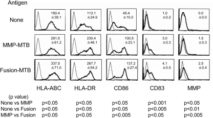
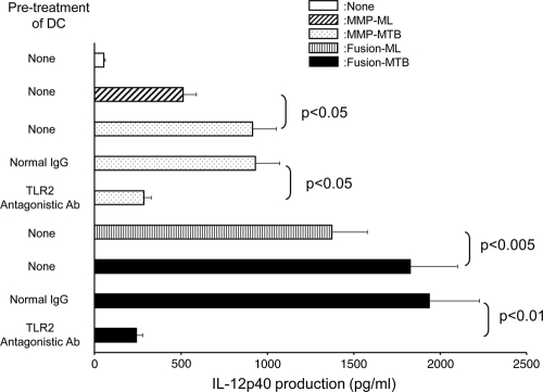
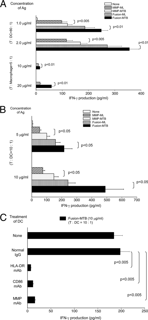
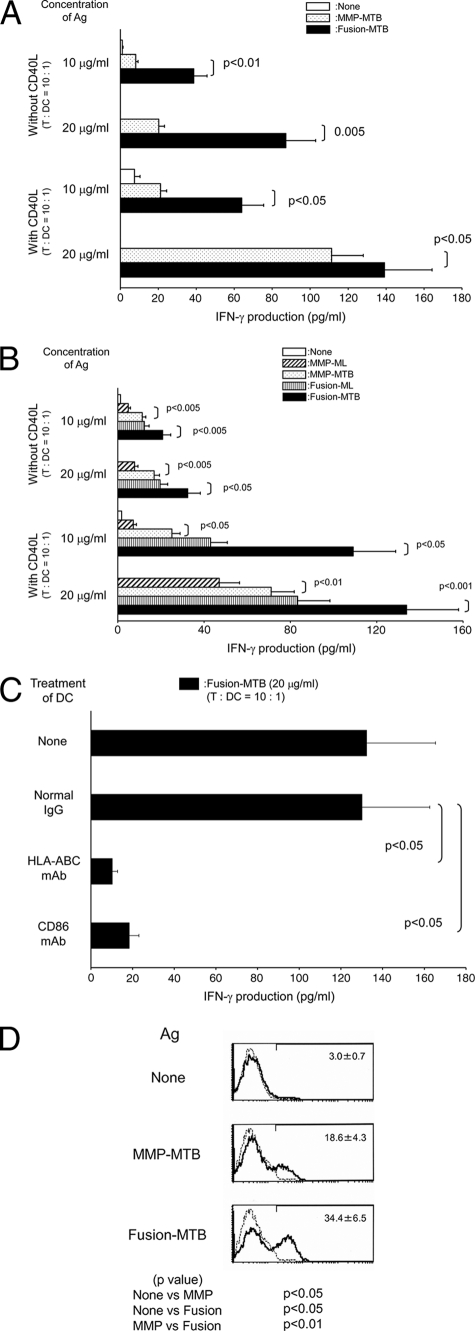
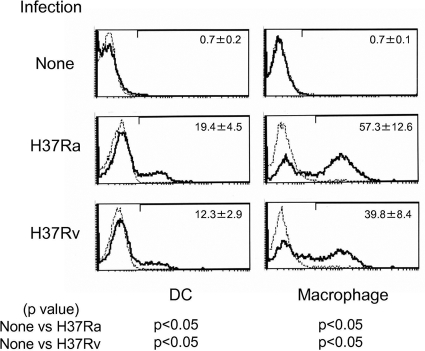
Previously, we observed that both major membrane protein II of Mycobacterium leprae (MMP-ML) and its fusion with M. bovis BCG (BCG)-derived heat shock protein 70 (HSP70) (Fusion-ML) are immunogenic and that recombinant BCG secreting either of these proteins effectively inhibits the multiplication of M. leprae in mice. Here, we purified M. tuberculosis-derived major membrane protein II (MMP-MTB) and its fusion with HSP70 (Fusion-MTB) in a lipopolysaccharide-free condition and evaluated their immunostimulatory abilities. Both MMP-MTB and Fusion-MTB activated monocyte-derived dendritic cells (DC) in terms of phenotype and interleukin-12 (IL-12) production, but Fusion-MTB more efficiently activated them than MMP-MTB did. The IL-12 production was a consequence of the ligation of those recombinant proteins with Toll-like receptor 2. The M. tuberculosis-derived and M. leprae-derived recombinant proteins activated naïve T cells of both CD4 and CD8 subsets, but M. tuberculosis-derived proteins were superior to M. leprae-derived proteins and fusion proteins were superior to MMP, regardless of the origin of the protein. Memory-type CD4(+) T cells obtained from BCG-vaccinated healthy individuals seem to be primed with MMP-MTB by the vaccination, and both M. tuberculosis-derived recombinant proteins produced perforin-producing CD8(+) T cells from memory-type CD8(+) T cells. Further, infection of DC and macrophages with M. tuberculosis H37Ra and H37Rv induced the expression of MMP on their surface. These results indicate that M. tuberculosis-derived MMP, as a sole protein or as part of a fusion protein, may be useful for developing new vaccinating agents against tuberculosis.
Figures
Similar articles
-
Polyclonal activation of naïve T cells by urease deficient-recombinant BCG that produced protein complex composed of heat shock protein 70, CysO and major membrane protein-II.BMC Infect Dis. 2014 Apr 2;14:179. doi: 10.1186/1471-2334-14-179. BMC Infect Dis. 2014. PMID: 24690183 Free PMC article.
-
Efficient activation of human T cells of both CD4 and CD8 subsets by urease-deficient recombinant Mycobacterium bovis BCG that produced a heat shock protein 70-M. tuberculosis-derived major membrane protein II fusion protein.Clin Vaccine Immunol. 2014 Jan;21(1):1-11. doi: 10.1128/CVI.00564-13. Epub 2013 Oct 23. Clin Vaccine Immunol. 2014. PMID: 24152387 Free PMC article.
-
Induction of cross-priming of naive CD8+ T lymphocytes by recombinant bacillus Calmette-Guerin that secretes heat shock protein 70-major membrane protein-II fusion protein.J Immunol. 2009 Nov 15;183(10):6561-8. doi: 10.4049/jimmunol.0803857. Epub 2009 Oct 21. J Immunol. 2009. PMID: 19846882
-
[Enhanced activation of T lymphocytes by urease-deficient recombinant bacillus Calmette-Guérin producing heat shock protein 70-major membrane protein-II fusion protein].Nihon Hansenbyo Gakkai Zasshi. 2012 Sep;81(3):199-203. doi: 10.5025/hansen.81.199. Nihon Hansenbyo Gakkai Zasshi. 2012. PMID: 23012848 Review. Japanese.
-
[Development of a novel recombinant BCG for tuberculosis vaccine].Nihon Hansenbyo Gakkai Zasshi. 2013 Dec;82(3):107-10. doi: 10.5025/hansen.82.107. Nihon Hansenbyo Gakkai Zasshi. 2013. PMID: 24579457 Review. Japanese.
Cited by
-
Polyclonal activation of naïve T cells by urease deficient-recombinant BCG that produced protein complex composed of heat shock protein 70, CysO and major membrane protein-II.BMC Infect Dis. 2014 Apr 2;14:179. doi: 10.1186/1471-2334-14-179. BMC Infect Dis. 2014. PMID: 24690183 Free PMC article.
-
Diagnostic performance of culture filtered protein 10-specific perforin in pediatric patients with active tuberculosis.J Clin Lab Anal. 2020 Nov;34(11):e23477. doi: 10.1002/jcla.23477. Epub 2020 Jul 16. J Clin Lab Anal. 2020. PMID: 32671908 Free PMC article.
-
Efficient activation of human T cells of both CD4 and CD8 subsets by urease-deficient recombinant Mycobacterium bovis BCG that produced a heat shock protein 70-M. tuberculosis-derived major membrane protein II fusion protein.Clin Vaccine Immunol. 2014 Jan;21(1):1-11. doi: 10.1128/CVI.00564-13. Epub 2013 Oct 23. Clin Vaccine Immunol. 2014. PMID: 24152387 Free PMC article.
-
Antigen-specific T cells and cytokines detection as useful tool for understanding immunity against zoonotic infections.Clin Dev Immunol. 2012;2012:768789. doi: 10.1155/2012/768789. Epub 2012 Feb 9. Clin Dev Immunol. 2012. PMID: 22400039 Free PMC article. Review.
-
New findings of Toll-like receptors involved in Mycobacterium tuberculosis infection.Pathog Glob Health. 2017 Jul;111(5):256-264. doi: 10.1080/20477724.2017.1351080. Epub 2017 Jul 17. Pathog Glob Health. 2017. PMID: 28715935 Free PMC article. Review.
References
-
- Aagaard, C. S., T. T. K. T. Hoang, C. Vingsbo-Lundberg, J. Dietrich, and P. Andersen. 2009. Quality and vaccine efficacy of CD4+ T cell responses directed to dominant and subdominant epitopes in ESAT-6 from Mycobacterium tuberculosis. J. Immunol. 183:2659-2668. - PubMed
-
- Andersen, P. 2007. Tuberculosis vaccines: an update. Nat. Rev. Microbiol. 5:484-487. - PubMed
-
- Andersen, P., and T. M. Doherty. 2005. The success and failure of BCG: implications for a novel tuberculosis vaccine. Nat. Rev. Microbiol. 3:656-662. - PubMed
-
- Bardarov, S., et al. 2002. Specialized transduction: an efficient method for generating marked and unmarked targeted gene disruptions in Mycobacterium tuberculosis, M. bovis BCG and M. smegmatis. Microbiology 148(Pt. 10):3007-3017. - PubMed
Publication types
MeSH terms
Substances
LinkOut - more resources
Full Text Sources
Research Materials
