. 2010 Dec 15;30(50):16910-21.
doi: 10.1523/JNEUROSCI.3660-10.2010.

Fragile X protein FMRP is required for homeostatic plasticity and regulation of synaptic strength by retinoic acid

Affiliations

Fragile X protein FMRP is required for homeostatic plasticity and regulation of synaptic strength by retinoic acid

Marta E Soden et al. J Neurosci. .

Abstract

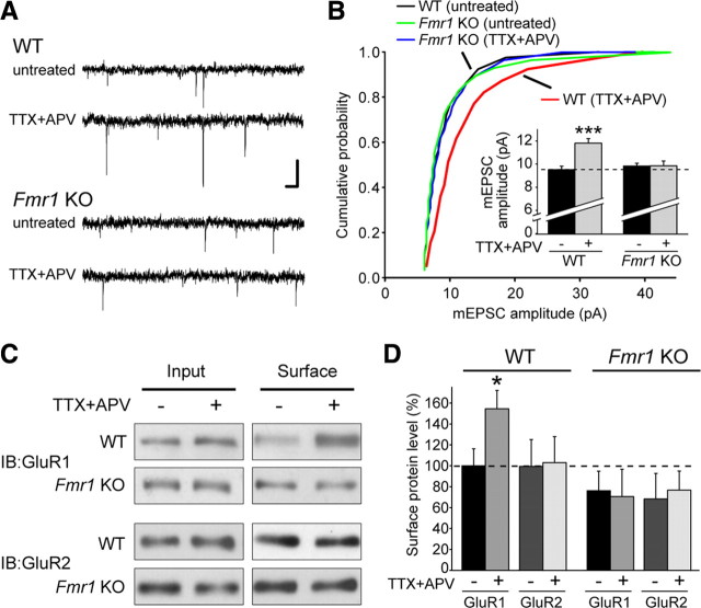
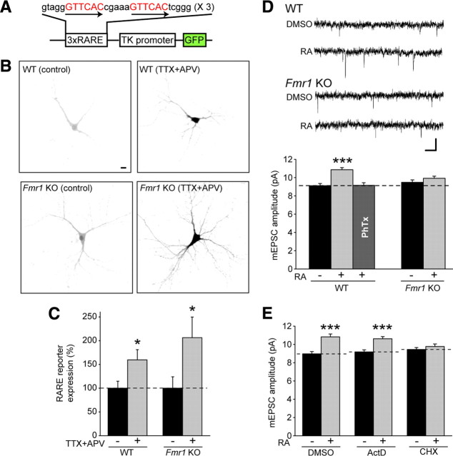
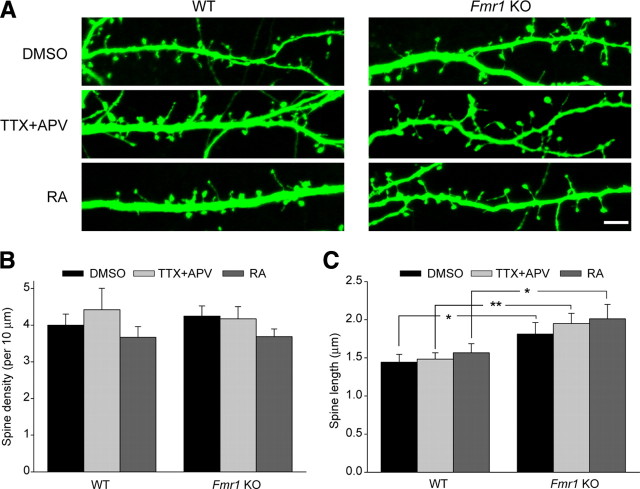
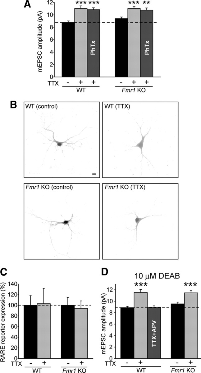
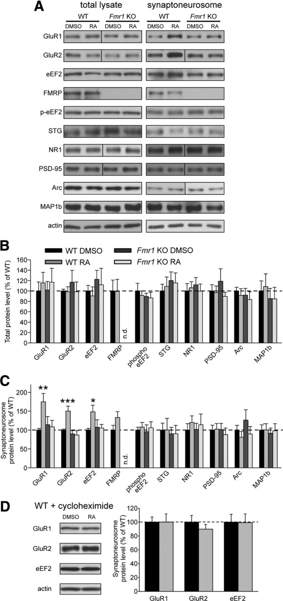
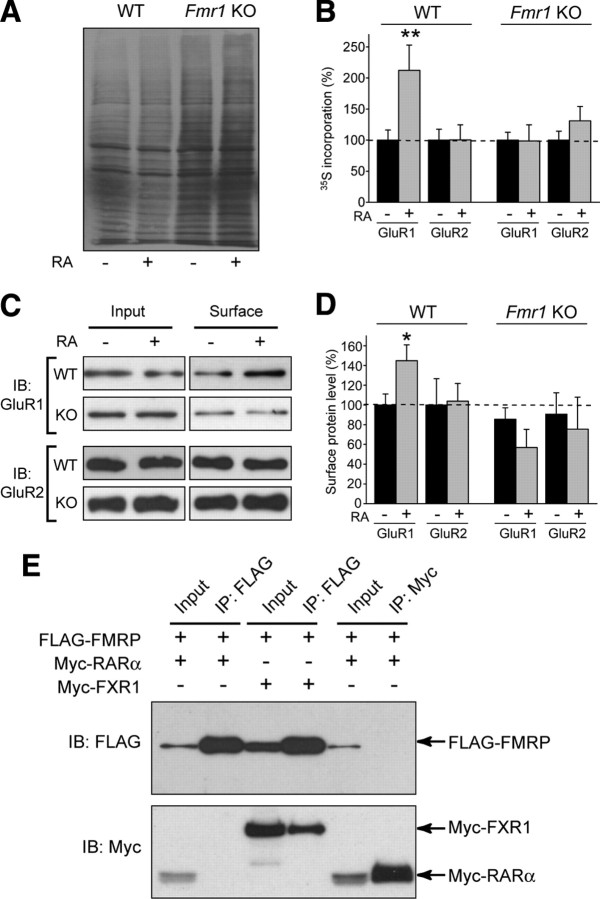
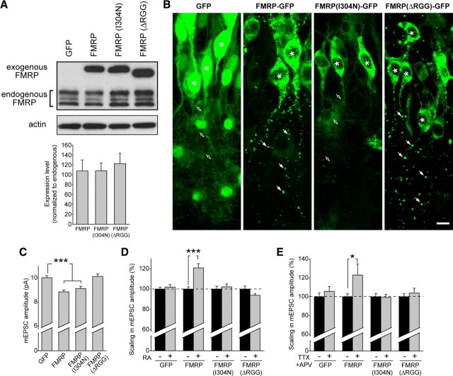
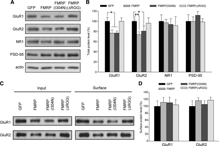
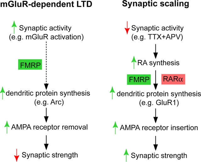
Homeostatic synaptic plasticity adjusts the strength of synapses during global changes in neural activity, thereby stabilizing the overall activity of neural networks. Suppression of synaptic activity increases synaptic strength by inducing synthesis of retinoic acid (RA), which activates postsynaptic synthesis of AMPA-type glutamate receptors (AMPARs) in dendrites and promotes synaptic insertion of newly synthesized AMPARs. Here, we show that fragile X mental retardation protein (FMRP), an RNA-binding protein that regulates dendritic protein synthesis, is essential for increases in synaptic strength induced by RA or by blockade of neural activity in the mouse hippocampus. Although activity-dependent RA synthesis is maintained in Fmr1 knock-out neurons, RA-dependent dendritic translation of GluR1-type AMPA receptors is impaired. Intriguingly, FMRP is only required for the form of homeostatic plasticity that is dependent on both RA signaling and local protein synthesis. Postsynaptic expression of wild-type or mutant FMRP(I304N) in knock-out neurons reduced the total, surface, and synaptic levels of AMPARs, implying a role for FMRP in regulating AMPAR abundance. Expression of FMRP lacking the RGG box RNA-binding domain had no effect on AMPAR levels. Importantly, postsynaptic expression of wild-type FMRP, but not FMRP(I304N) or FMRPΔRGG, restored synaptic scaling when expressed in knock-out neurons. Together, these findings identify an unanticipated role for FMRP in regulating homeostatic synaptic plasticity downstream of RA. Our results raise the possibility that at least some of the symptoms of fragile X syndrome reflect impaired homeostatic plasticity and impaired RA signaling.


Figures

Figure 1.
Figure 1.
FMRP is required for TTX + APV-induced synaptic scaling. A, Representative mEPSC traces from wild-type and Fmr1 knock-out (untreated and TTX + APV treated) neurons in hippocampal slice culture. Calibration: 10 pA, 40 ms. B, Cumulative distribution of mEPSC amplitudes from WT and KO neurons treated with 36 h of TTX + APV (p < 0.001, Kolmogorov–Smirnov test). Inset, Quantification of average mEPSC amplitude (n = 28–34; ***p < 0.001). C, Representative blots for biotinylation of surface AMPARs in primary cultured neurons after 24 h of TTX + APV treatment. IB, Immunoblot. D, Quantification of C. Surface band intensity was normalized to input, and all groups were compared with WT untreated (n = 4–6; *p < 0.05). Error bars represent SEM.
Figure 2.
Figure 2.
FMRP is not required for RA synthesis but is specifically required for RA-induced local translation-dependent synaptic scaling. A, Schematic of the 3xDR5–RARE–GFP reporter construct. B, Representative images of RARE–GFP reporter expression in WT and KO neurons with and without 24 h TTX + APV treatment. Scale bar, 10 μm. C, Quantification of B (n = 16–18; *p < 0.05). D, Representative traces and quantification of mEPSC amplitude in WT and KO neurons after 4 h DMSO or RA treatment (n = 31–33; ***p < 0.001). Philanthotoxin-433 was used to block GluR2-lacking AMPA receptor-mediated responses in the WT–RA group (n = 22). Calibration: 10 pA, 40 ms. E, Effect of transcription inhibitor actinomycin D and translation inhibitor cycloheximide on RA-induced synaptic scaling in WT neurons (n = 22–27; ***p < 0.001). Error bars represent SEM.
Figure 3.
Figure 3.
RA does not affect spine morphology in WT or KO neurons. A, Sample images of GFP-expressing WT or KO neurons treated with TTX + APV or RA. Scale bar, 5 μm. B, C, Quantification of spine density and spine length in WT and KO neurons treated with 24 h of TTX + APV or 30 min (plus 1 h washout) of RA (n = 9–10 cells per group, 2–3 branches per cell). For spine density, p > 0.5. For spine length *p < 0.05, **p < 0.01. Error bars represent SEM.
Figure 4.
Figure 4.
Neither FMRP nor RA is required for TTX-alone-induced synaptic scaling. A, Sixty hours of TTX induced synaptic scaling in WT and KO neurons. PhTx was used to block GluR2-lacking AMPA receptor-mediated responses (n = 18–39; **p < 0.01; ***p < 0.001). B, Representative images of RARE–GFP reporter expression in WT and KO neurons with and without 48 h of TTX treatment. Scale bar, 10 μm. C, Quantification of B (n = 22–28). D, Effect of the RA synthesis blocker DEAB on 60 h of TTX-induced synaptic scaling and 36 h of TTX + APV-induced scaling (n = 21–27; ***p < 0.001). Error bars represent SEM.
Figure 5.
Figure 5.
FMRP is required for RA-induced local translation of specific synaptic proteins. A, Representative blots of synaptic proteins from whole lysates and synaptoneurosome (SNS) fractions of WT and KO hippocampal slices, treated with 4 h of DMSO or RA. A vertical line indicates the removal (for ease of viewing) of extra lanes between WT and KO lanes. B, C, Quantification of synaptic proteins in the total lysate (B) and synaptoneurosome fraction (C) from treated hippocampal slices. Band intensities were normalized to actin (n.d., not detected; STG, Stargazin; n = 5–16; *p < 0.05; **p < 0.01; ***p < 0.001). D, Representative blots and quantification of proteins from the synaptic fraction of WT slices pretreated with the translation blocker cycloheximide before DMSO or RA treatment (n = 5). Error bars represent SEM.
Figure 6.
Figure 6.
Metabolic labeling shows increased GluR1 translation with RA treatment, and FMRP and RARα do not interact directly. A, Autoradiography of total protein lysate from dissociated neurons treated with 2 h of DMSO or RA in the presence of 35S-labeled amino acids. B, Quantification of incorporated radioactivity using liquid scintillation counting after immunoprecipitation of AMPARs. Each genotype was normalized separately to its DMSO group (n = 5–8; **p < 0.01). C, Representative blots for biotinylation of surface AMPARs in primary cultured neurons after RA treatment. IB, Immunoblot. D, Quantification of surface GluR1 and GluR2 protein levels after RA treatment. Surface band intensity was normalized to input, and all groups were compared with WT DMSO (n = 3–9; *p < 0.05). E, Attempted coimmunoprecipitation of FMRP and RARα. Tagged constructs were expressed in HEK293T cells as indicated, and immunoprecipitation (IP) of either FMRP (anti-FLAG) or RARα (anti-Myc) was performed (Input: 10% of total). Although pull down of FMRP was able to coimmunoprecipitate the known binding partner FXR1 (positive control), no interaction was seen between FMRP and RARα. Error bars represent SEM.
Figure 7.
Figure 7.
Viral expression of FMRP in knock-out slices restores synaptic scaling. A, Representative blot and quantification of exogenous FMRP–GFP, FMRP(I304N)–GFP, and FMRPΔRGG–GFP expression compared with endogenous FMRP protein levels after 6 d of virus expression in wild-type dissociated neurons (n = 6). B, Images from the CA1 region of hippocampal slices infected with lentiviral constructs expressing GFP, FMRP–GFP, FMRP(I304N)–GFP, or FMRPΔRGG–GFP. Asterisks indicate cell body. FMRP and FMRPΔRGG exhibit a punctate expression pattern in neuronal dendrites (filled arrows), whereas GFP or FMRP(I304N) are diffusely expressed in neuronal dendrites (open arrows). Scale bar, 10 μm. C, Amplitude of mEPSC events in KO neurons expressing GFP, FMRP, FMRP(I304N), or FMRPΔRGG (n = 38–66; ***p < 0.001). D, Percentage scaling after RA treatment in neurons expressing GFP or different FMRP constructs. DMSO groups for each construct were set to 100% to account for altered baseline amplitudes (see C) (n = 18–49; ***p < 0.001). E, Percentage scaling after treatment with 36 h of TTX + APV (n = 17–20; *p < 0.05). Error bars represent SEM.
Figure 8.
Figure 8.
Viral expression of FMRP in knock-out neurons affects AMPAR abundance. Representative blots (A) and quantification of protein levels in lysates (B) collected from KO dissociated neurons infected with virus expressing GFP, FMRP, FMRP(I304N), or FMRPΔRGG. Band intensities were normalized to actin (n = 4–10; *p < 0.05; **p < 0.01). Representative blots (C) and quantification of surface biotinylation of AMPARs (D) in knock-out dissociated neurons infected with virus expressing GFP or different FMRP constructs. Surface band intensity was normalized to input (n = 5–7). Error bars represent SEM.
Figure 9.
Figure 9.
FMRP plays a critical role in multiple forms of synaptic plasticity. Synaptic activation leads to FMRP-dependent protein synthesis and the eventual removal of AMPARs from the synapse (mGluR-dependent LTD). By contrast, blockade of synaptic activity causes synthesis of new GluR1 receptors and a subsequent increase in synaptic strength, in a process that requires both FMRP and RARα.

Comment in

References

    1. Antar LN, Afroz R, Dictenberg JB, Carroll RC, Bassell GJ. Metabotropic glutamate receptor activation regulates fragile x mental retardation protein and FMR1 mRNA localization differentially in dendrites and at synapses. J Neurosci. 2004;24:2648–2655. - PMC - PubMed
    1. Aoto J, Nam CI, Poon MM, Ting P, Chen L. Synaptic signaling by all-trans retinoic acid in homeostatic synaptic plasticity. Neuron. 2008;60:308–320. - PMC - PubMed
    1. Aschrafi A, Cunningham BA, Edelman GM, Vanderklish PW. The fragile X mental retardation protein and group I metabotropic glutamate receptors regulate levels of mRNA granules in brain. Proc Natl Acad Sci U S A. 2005;102:2180–2185. - PMC - PubMed
    1. Ashley CT, Jr, Wilkinson KD, Reines D, Warren ST. FMR1 protein: conserved RNP family domains and selective RNA binding. Science. 1993;262:563–566. - PubMed
    1. Bassell GJ, Warren ST. Fragile X syndrome: loss of local mRNA regulation alters synaptic development and function. Neuron. 2008;60:201–214. - PMC - PubMed

Publication types

MeSH terms

LinkOut - more resources