. 2010 Dec 15;30(50):16922-37.
doi: 10.1523/JNEUROSCI.3644-10.2010.

Projection-specific neuromodulation of medial prefrontal cortex neurons

Affiliations

Projection-specific neuromodulation of medial prefrontal cortex neurons

Nikolai C Dembrow et al. J Neurosci. .

Abstract

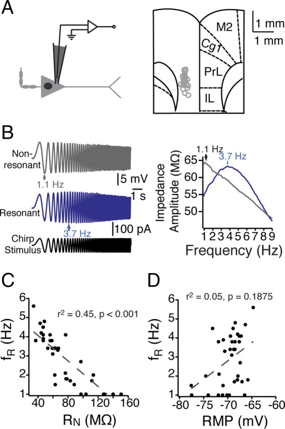
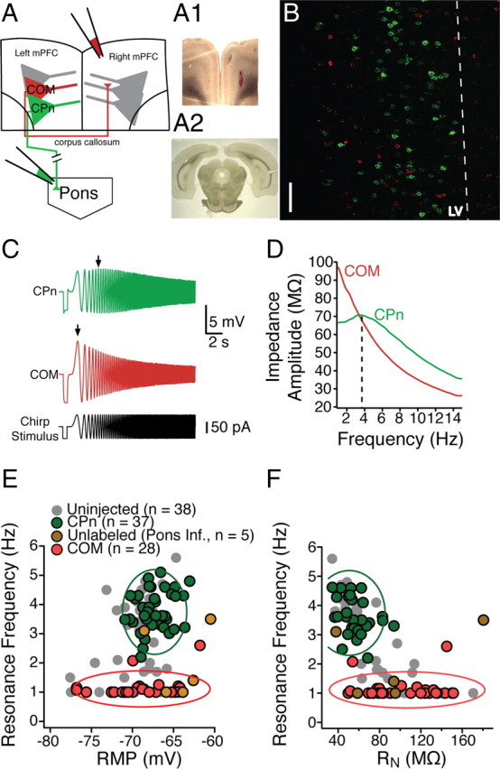
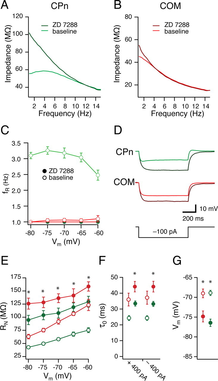
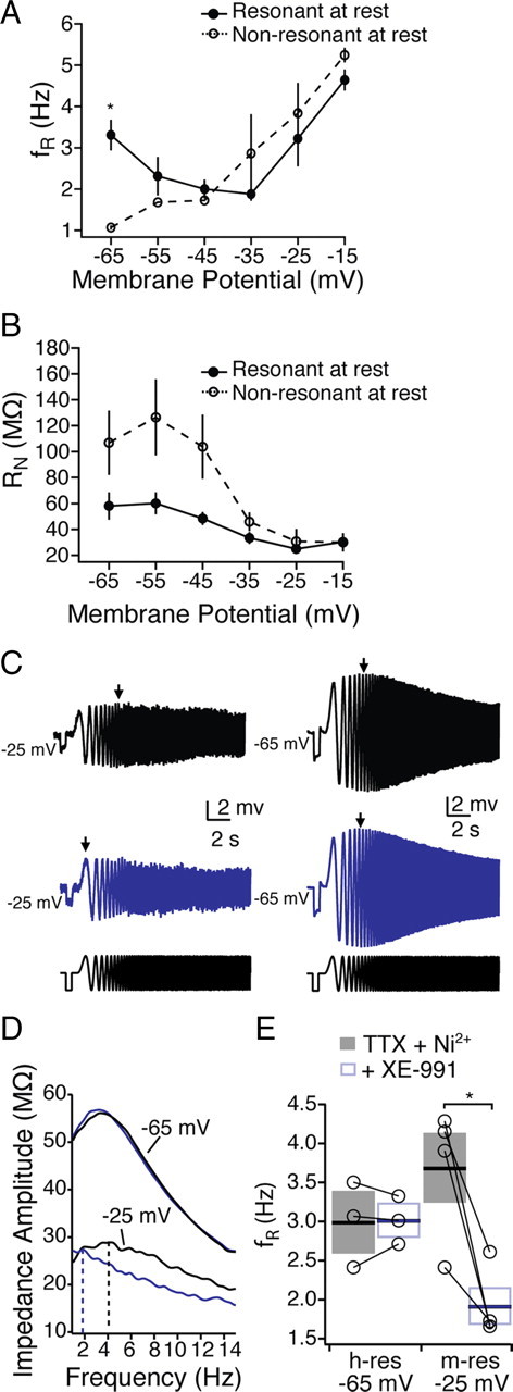
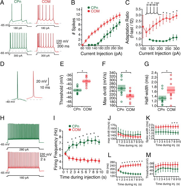
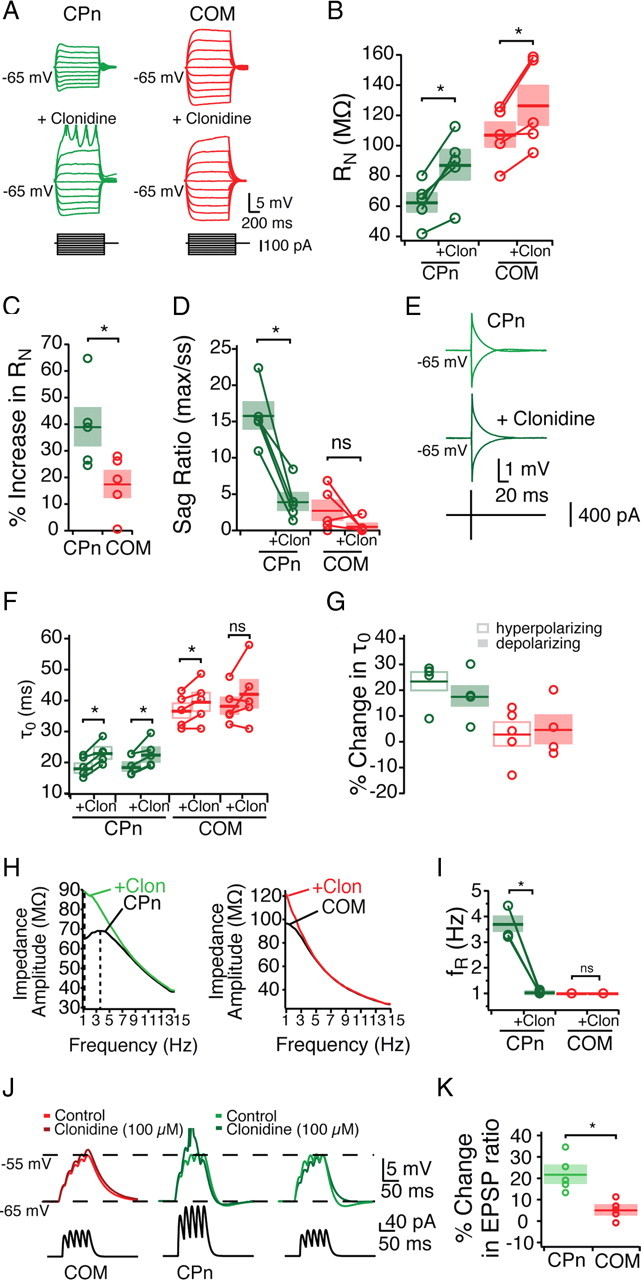
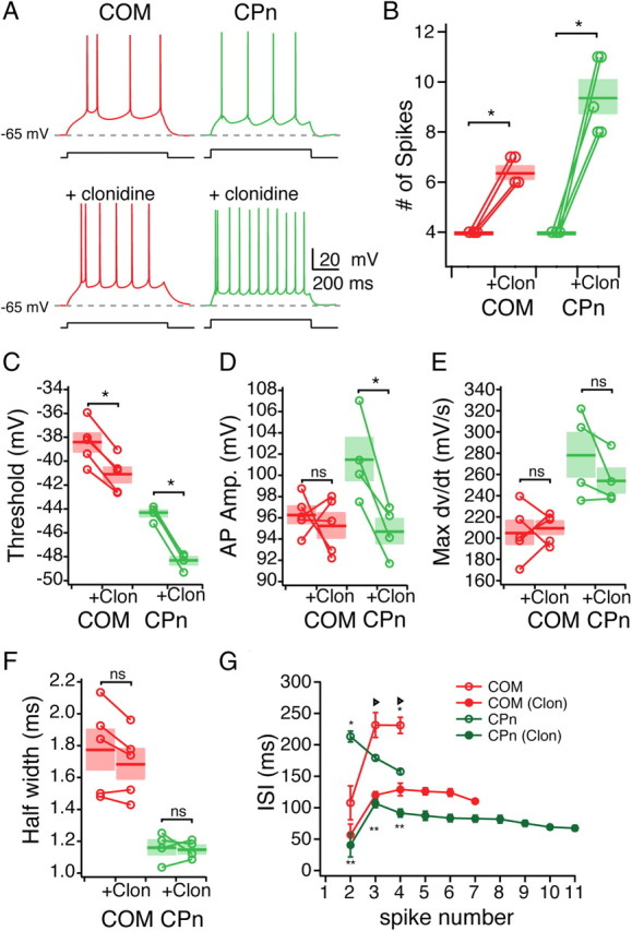
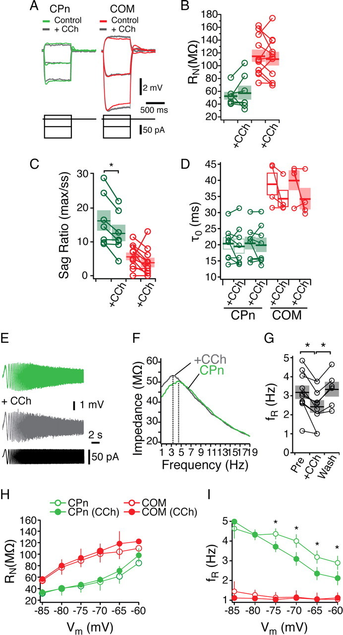
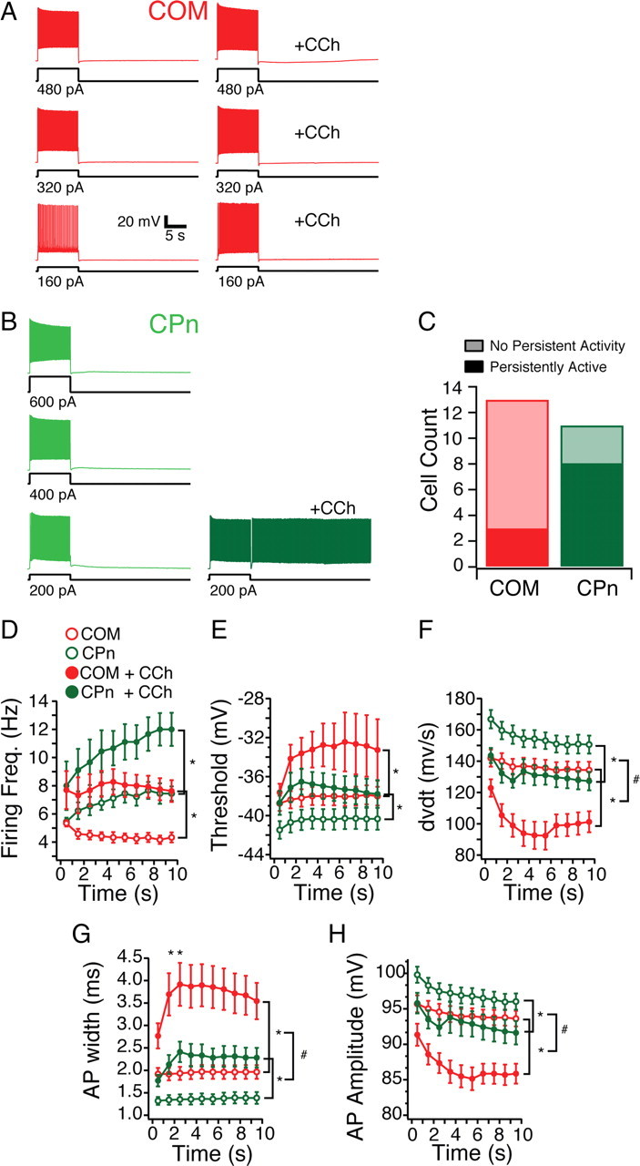
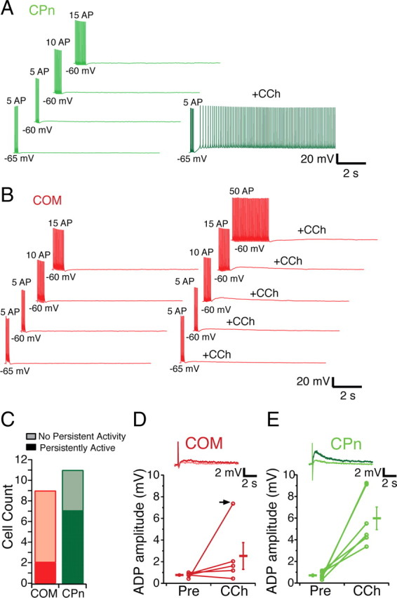
Mnemonic persistent activity in the prefrontal cortex (PFC) constitutes the neural basis of working memory. To understand how neuromodulators contribute to the generation of persistent activity, it is necessary to identify the intrinsic properties of the layer V pyramidal neurons that transfer this information to downstream networks. Here we show that the somatic dynamic and integrative properties of layer V pyramidal neurons in the rat medial PFC depend on whether they project subcortically to the pons [corticopontine (CPn)] or to the contralateral cortex [commissural (COM)]. CPn neurons display low temporal summation and accelerate in firing frequency when depolarized, whereas COM neurons have high temporal summation and display spike frequency accommodation. In response to dynamic stimuli, COM neurons act as low-pass filters, whereas CPn neurons act as bandpass filters, resonating in the theta frequency range (3-6 Hz). The disparate subthreshold properties of COM and CPn neurons can be accounted for by differences in the hyperpolarization-activated cyclic nucleotide gated cation h-current. Interestingly, neuromodulators hypothesized to enhance mnemonic persistent activity affect COM and CPn neurons distinctly. Adrenergic modulation shifts the dynamic properties of CPn but not COM neurons and increases the excitability of CPn neurons significantly more than COM neurons. In response to cholinergic modulation, CPn neurons were much more likely to display activity-dependent intrinsic persistent firing than COM neurons. Together, these data suggest that the two categories of projection neurons may subserve separate functions in PFC and may be engaged differently during working memory processes.


Figures

Figure 1.
Figure 1.
Heterogeneity in the dynamic properties of mPFC neurons. A, Somatic recordings of layer V mPFC neurons were conducted within ventral mPFC, including prelimbic and infralimbic cortex. Left, A schematic of an mPFC neuron and the somatic recording location. Right, Recording locations overlaid with a modified version of a coronal diagram from a rat brain atlas (Paxinos and Watson, 1993). B, In response to a 10 s chirp stimulus at their resting membrane potential, different mPFC neurons resonated across a range (1–6 Hz) of frequencies. C, D, In addition to exhibiting different resonance profiles, neurons were diverse in both steady-state input resistance (C) and resting membrane potential (D) (n = 38 neurons). Gray dashed lines represent the linear fit of the data, with correlation values listed. PL, Prelimbic; IL, infralimbic; Cg1, anterior cingulate; M2, secondary motor cortices.
Figure 2.
Figure 2.
Dynamics of mPFC neurons depend on their long-range projection targets. A, Schematic of dual infusion strategy. Retrograde beads were infused into contralateral mPFC and/or the ipsilateral pyramidal tracts/pons. A1, Coronal slice, 300 μm thick, containing the contralateral mPFC infusion site. A2, Ipsilateral pyramidal tract infusion site in a 50 μm coronal section. B, Distribution of COM and CPn neurons. COM and CPn neurons are two nonoverlapping populations. CPn neurons (green) form a band at the upper parts of layer V (LV). COM neurons (red) are interspersed throughout layer V in close proximity to CPn neurons as well as in more superficial layers. Scale bar, 50 μm. C, In response to a 15 s, 15 Hz chirp stimulus, CPn neurons (green) resonate at a much higher frequency than COM neurons (red). D, A comparison of the ZAP of COM versus CPn neurons. E, F, Overlay of the resonance profiles plotted against membrane potential (E) and input resistance (F). Neurons were labeled by the pyramidal tract/pontine infusion (green), neurons did not get labeled by the pontine pyramidal tract/pontine infusion (gold), or COM neurons labeled from the contralateral infusion (red). For comparison, unlabeled neurons from Figure 1, C and D, are shown in gray. Cluster boundaries (solid lines) were found using mean cluster analysis and represent the SD around each cluster.
Figure 3.
Figure 3.
Representative morphologies of mPFC COM and CPn neurons. A, Representative reconstructions from Neurobiotin filled and subsequently DAB processed identified COM (red) and CPn (Green) neurons. B, C, Sholl analysis (grouped into 30 μm segments) illustrates the distinct morphology of COM (n = 5) versus CPn (n = 5) neurons in the apical tuft region. D–F, Dendritic lengths (D), surface area (E), and volume (F) from the two neuron types. *p < 0.05.
Figure 4.
Figure 4.
Subthreshold properties of CPn and COM neurons. Somatic patch recordings of labeled COM and CPn neurons reveal that they have distinct subthreshold properties. A, CPn neurons are slightly more depolarized than COM neurons, although not significantly. For the purpose of comparison, neurons were held at −65 mV during stimulus protocols. B–F, COM neurons (red) have higher steady-state input resistance (B), lower sag ratio (C), slower time constant in both the hyperpolarizing and depolarizing directions (D), less rebound (E), and more temporal summation (F) than CPn neurons (green). *p < 0.05.
Figure 5.
Figure 5.
Differences in h-conductance contribute to distinct dynamic and steady-state properties. A, B, COM (red) and CPn (green) neurons were given 15 s, 15 Hz chirp stimuli across a range of membrane potentials (C; −80 to −60 mV) before and after bath application of 20 μm ZD7288, eliminating membrane resonance in CPn neurons. Representative ZAPs at −70 mV. A, B, Steady-state properties were affected in both groups. D, Representative traces of a single current step (−100 pA) in CPn (green) and COM (red) neurons before and after the addition of ZD7288 (black). E, Input resistance measured as the slope of the linear fit from a family of current injections in CPn (green) and COM (red) neurons before (open circles) and after (filled circles) the addition of ZD7288. F, G, ZD7288 (filled circles) changes both the functional membrane time constant at −65 mV (F) and the resting membrane potential (G) in both CPn (green) and COM (red) neurons. *p < 0.05.
Figure 6.
Figure 6.
m-resonance in mPFC neurons. In the presence of TTX and Ni2+, mPFC neurons were segregated into resonant and nonresonant depending on their resonance at −65 mV. A, B, Resonance frequency (A) and input resistance (B) from resonant and nonresonant neurons from −65 to −25 mV. C–E, The m-channel blocker XE991 (10 μm) blocked resonance at −25 mV, whereas resonance at −65 mV was unaffected. *p < 0.05.
Figure 7.
Figure 7.
Active properties of COM versus CPn neurons. A, Representative responses of COM (red) and CPn neuron responses to 750 ms step depolarizations of 180 and 300 pA. B, Firing frequency of spikes elicited across a range of current injections (60–300 pA). C, Ratio of the first ISI to the last ISI. D, Representative traces of the first action potential in COM and CPn neurons with sufficient current to trigger four action potentials in 750 ms. Action potentials in CPn neurons were typically followed by a fast afterhyperpolarization and fast ADP. E–G, Threshold, rate of rise (max dv/dt), and AP half-widths of COM and CPn neurons. H, COM neurons exhibit spike frequency adaptation, whereas CPn neurons exhibit spike acceleration. Representative traces of a 10 s current injection that elicits 5 Hz firing frequency in the first second. I–M, Changes in firing frequency, max dv/dt, interspike interval, spike width, and threshold over the course of a 10 s current injection. *p < 0.05.
Figure 8.
Figure 8.
Noradrenergic modulation of subthreshold properties of mPFC neurons. Bath application of the α2-adrenergic agonist clonidine (Clon; 100 μm) differentially effects CPn and COM neurons. A–G, Effects of clonidine on steady-state input resistance (A–C), sag ratio (D), and membrane time constant (E–G). H, I, Clonidine shifts the ZAP of CPn but not COM neurons, making CPn neurons drop in resonance frequency. J, αEPSPs were injected with current such that they depolarized the neurons from −65 to −55 mV. With the addition of clonidine, only CPn neurons are driven to fire an action potential. K, Reducing the current injection to span from −65 to −55 mV in the presence of clonidine reveals an increase in CPn but not COM EPSP ratio. *p < 0.05.
Figure 9.
Figure 9.
Noradrenergic modulation of the excitability of mPFC neurons. A, Representative traces of the effect of clonidine (100 μm) on the excitability of COM (Red) and CPn (green) neurons. B, Group data showing the increase in the number of spikes elicited in COM and CPn neurons before and after the addition of clonidine (Clon). C–F, Changes in spike parameters caused by clonidine in COM and CPn neurons. G, Changes in the interspike interval of both COM and CPn neurons. *p < 0.05 (significant difference between COM and CPN in control conditions); **p < 0.05 (significant difference between CPn control and CPn clonidine); triangle, p < 0.05 (significant difference between COM control and COM clonidine).
Figure 10.
Figure 10.
Cholinergic modulation of subthreshold properties in mPFC neurons. A, Representative traces current injections in COM (red) and CPn (green) neurons. Overlays are with the addition of 20 μm the cholinergic agonist CCh (gray). B–D, Changes in steady-state input resistance (B), sag ratio (C), and membrane time constant (D) elicited by CCh. E–G, CCh reduced the resonance frequency of CPn neurons in a reversible manner. H, I, Input resistance (H) and resonance frequency (I) before (open circles) and in the presence of 20 μm CCh (filled circles) in both COM (red) and CPn (green) neurons at holding potentials from −85 to −60 mV. *p < 0.05.
Figure 11.
Figure 11.
Projection dependence of single-neuron persistent activity in the presence of cholinergic modulation. A, COM neurons are depolarized for 10 s with one, two, and three times the amount of current sufficient to drive 5 Hz firing frequency for the initial second. With the addition of the cholinergic agonist (CCh, 20 μm), COM neurons exhibit a slight afterdepolarizing potential but no persistent activity. B, CPn neurons depolarized in the same manner fire persistently only with the addition of CCh with one time the current injection. C, A greater proportion of CPn (green) neurons could be triggered to fire persistently in the presence of CCh by 10 s depolarization than COM neurons (red). D–I, Spiking characteristics of COM (red) and CPn (green) neurons before (open circles) and with the addition of 20 μm CCh (filled circles). *p < 0.05 (significant difference between control and CCh condition); #p < 0.05 (significant difference in the effect of CCh on COM vs CPn neurons).
Figure 12.
Figure 12.
Subcortically projecting neurons can be driven to fire persistently with spike trains. A, CPn neurons are driven to fire 5, 10, or 15 action potentials with 1 ms, 20 Hz trains of depolarizing current from either −65 or −60 mV. In the presence of 20 μm CCh, these trains are sufficient to cause CPn neurons to persistently fire. B, COM neurons cannot be driven to fire persistently in the presence of CCh even with 50 APs delivered in the same manner. C, Counts of COM (red) and CPn (green) neurons that fire persistently using trains of spikes. D, E, In both COM (light red, inset) and CPn (light green, inset) neurons, a 1 nA, 10 ms depolarizing current injection does result in a small (0–1.5 mV) ADP. With the addition of 20 μm CCh, most COM neurons (4 of 5) exhibit a slight increase in the ADP (dark red, inset). In 1 of 5 COM neurons, a large ADP occurred with CCh (arrow). In CPn neurons (dark green, inset), a much larger ADP is elicited in the presence of CCh.

References

    1. Andrade R. Cell excitation enhances muscarinic cholinergic responses in rat association cortex. Brain Res. 1991;548:81–93. - PubMed
    1. Arnsten AF. Adrenergic targets for the treatment of cognitive deficits in schizophrenia. Psychopharmacology (Berl) 2004;174:25–31. - PubMed
    1. Arnsten AF. Stimulants: therapeutic actions in ADHD. Neuropsychopharmacology. 2006;31:2376–2383. - PubMed
    1. Arnsten AFT. Catecholamine and second messenger influences on prefrontal cortical networks of “representational knowledge”: a rational bridge between genetics and the symptoms of mental illness. Cereb Cortex. 2007;17(Suppl 1):i6–i15. - PubMed
    1. Barth AM, Vizi ES, Zelles T, Lendvai B. 2-Adrenergic receptors modify dendritic spike generation via HCN channels in the prefrontal cortex. J Neurophysiol. 2008;99:394–401. - PubMed

Publication types

LinkOut - more resources