. 2010 Dec 15;30(50):16938-48.
doi: 10.1523/JNEUROSCI.1598-10.2010.

Neuronal apoptosis induced by endoplasmic reticulum stress is regulated by ATF4-CHOP-mediated induction of the Bcl-2 homology 3-only member PUMA

Affiliations

Neuronal apoptosis induced by endoplasmic reticulum stress is regulated by ATF4-CHOP-mediated induction of the Bcl-2 homology 3-only member PUMA

Zohreh Galehdar et al. J Neurosci. .

Abstract

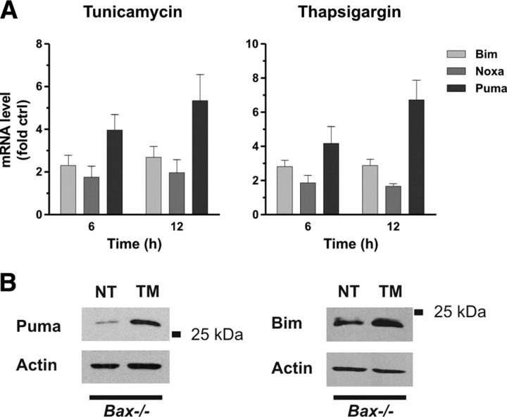
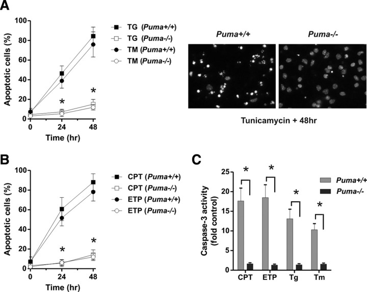
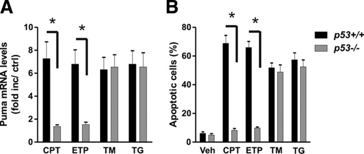
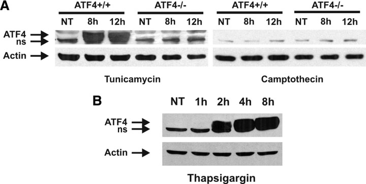
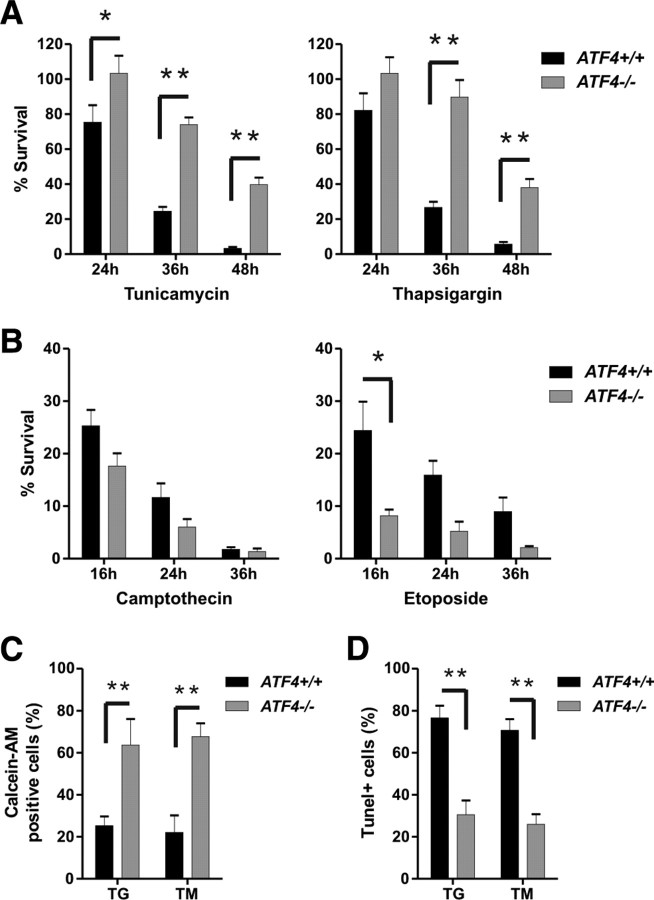
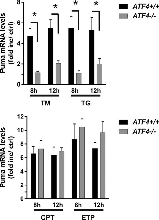
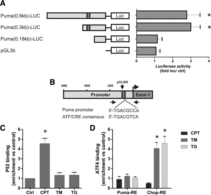
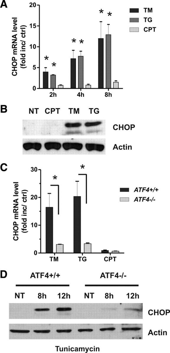
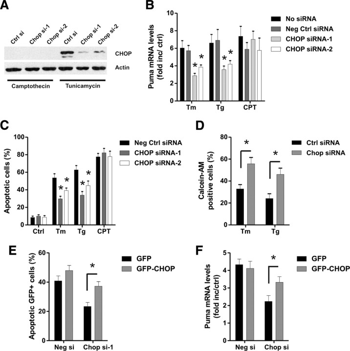
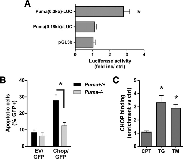
An increasing body of evidence points to a key role of endoplasmic reticulum (ER) stress in acute and chronic neurodegenerative conditions. Extensive ER stress can trigger neuronal apoptosis, but the signaling pathways that regulate this cell death remain unclear. In the present study, we demonstrate that PUMA, a Bcl-2 homology 3 (BH3)-only member of the Bcl-2 family, is transcriptionally activated in cortical neurons by ER stress and is essential for ER-stress-induced cell death. PUMA is known to be a key transcriptional target of p53, but we have found that ER stress triggers PUMA induction and cell death through a p53-independent mechanism mediated by the ER-stress-inducible transcription factor ATF4 (activating transcription factor 4). Specifically, we demonstrate that ectopic expression of ATF4 sensitizes mouse cortical neurons to ER-stress-induced apoptosis and that ATF4-deficient neurons exhibit markedly reduced levels of PUMA expression and cell death. However, chromatin immunoprecipitation experiments suggest that ATF4 does not directly regulate the PUMA promoter. Rather, we found that ATF4 induces expression of the transcription factor CHOP (C/EBP homologous protein) and that CHOP in turn activates PUMA induction. Specifically, we demonstrate that CHOP binds to the PUMA promoter during ER stress and that CHOP knockdown attenuates PUMA induction and neuronal apoptosis. In summary, we have identified a key signaling pathway in ER-stress-induced neuronal death involving ATF4-CHOP-mediated transactivation of the proapoptotic Bcl-2 family member PUMA. We propose that this pathway may be an important therapeutic target relevant to a number of neurodegenerative conditions.


Figures

Figure 1.
Figure 1.
Puma expression is induced in cortical neurons during ER stress. A, Cortical neurons were treated with tunicamycin (3 μg/ml) or thapsigargin (1 μm), and Bim, Noxa, and Puma mRNA levels were assessed by real-time RT-PCR. Expression was normalized to ribosomal S12 levels and is reported as fold increase over untreated controls (n = 4). B, Bax-deficient cortical neurons were treated with tunicamycin (TM), and Puma and Bim protein levels were assessed by Western blot at 24 h. NT, Nontreated.
Figure 2.
Figure 2.
PUMA is required for DNA- damage-induced and ER-stress-induced apoptosis in cortical neurons. A, B, Cortical neurons derived from Puma wild-type and knock-out littermates were treated with 1 μm thapsigargin (TG, Tg), 3 μg/ml tunicamycin (TM, Tm), 10 μm camptothecin (CPT), or 10 μm etoposide (ETP), and the fraction of apoptotic cells was determined at 24 and 48 h by assessing nuclear morphology after Hoechst staining. The fraction of apoptotic neurons was significantly decreased in Puma+/+ versus Puma−/− cultures (n ≥ 7; *p < 0.001). C, Puma wild-type and knock-out cortical neurons were treated with DNA-damaging agents or ER stressors as above. Cell lysates obtained 20 h after treatment were assayed for caspase-3 activity (n = 5; *p < 0.001).
Figure 3.
Figure 3.
ER stress triggers Puma induction and neuronal apoptosis through a p53-independent mechanism. Cortical neurons derived from p53 wild-type and p53 knock-out littermates were treated with thapsigargin (1 μm; TG), tunicamycin (3 μg/ml; TM), camptothecin (10 μm; CPT), or etoposide (10 μm; ETP). A, RNA was extracted 12 h after treatment, and Puma mRNA levels were quantified by real-time RT-PCR. Puma expression was normalized to S12 levels and is reported as fold increase over untreated controls (n ≥ 6; *p < 0.01). B, Neurons were stained with Hoechst dye 30 h after treatment, and the fraction of apoptotic cells was determined by assessing nuclear morphology (n = 5; *p < 0.01). Veh, Vehicle.
Figure 4.
Figure 4.
ATF4 expression is induced by ER stress but not DNA damage. A, Cortical neurons derived from ATF4−/− mice and wild-type littermates were treated with tunicamycin (3 μg/ml) or camptothecin (10 μm), and ATF4 protein levels were assessed by Western blot. A nonspecific (ns) band was detected in extracts from both wild-type and ATF4-deficient neurons. NT, Nontreated. B, Cortical neurons were untreated or treated with thapsigargin (1 μm) for the indicated times, and ATF4 protein levels were assessed by Western blot.
Figure 5.
Figure 5.
Ectopic expression of ATF4 sensitizes neurons to ER-stress-induced but not DNA-damage-induced apoptosis. A, Cortical neurons were cotransfected with pGFP and either pcDNA3 [empty vector (EV)] or pcDNA3–ATF4, and, after 24 h, neurons were treated with camptothecin (10 μm), tunicamycin (2 μg/ml), or DMSO (0.1%) as a vehicle control. At the indicated times, neurons were fixed and stained with Hoechst 33258, and the fraction of apoptotic and nonapoptotic GFP-positive cells was assessed by fluorescence microscopy (n = 3; *p < 0.05). B, Cortical neurons were infected with recombinant adenovirus expressing GFP (Ad–GFP) or coexpressing ATF4 and GFP (Ad–ATF4/GFP) at 50 MOI and, after 48 h, treated with tunicamycin (2 μg/ml) or thapsigargin (1 μm). After 24 h, cells were fixed and stained with Hoechst 33258, and the fraction of apoptotic and nonapoptotic GFP-positive neurons was determined (n = 3; **p < 0.01).
Figure 6.
Figure 6.
ATF4-deficient neurons are protected against ER-stress-induced but not DNA-damage-induced apoptosis. A, B, Cortical neurons derived from ATF4−/− embryos and wild-type littermates were treated with the ER stressors tunicamycin (3 μg/ml) and thapsigargin (1 μm) or the DNA-damaging agents camptothecin (10 μm) and etoposide (10 μm). At the indicated times, cells were lysed, and the number of intact nuclei (viable cells) was counted. Survival was calculated as the ratio of viable drug-treated neurons to viable untreated neurons for each embryo (n ≥ 5; *p < 0.05, **p < 0.01). C, D, Wild-type and ATF4-deficient neurons were treated with thapsigargin (1 μm; TG) or tunicamycin (3 μg/ml; TM), and the percentage of calcein-AM-positive (live) cells and TUNEL-positive cells was measured after 36 h (n = 3; **p < 0.01).
Figure 7.
Figure 7.
ATF4 regulates Puma induction during ER stress but not DNA damage. Cortical neurons derived from ATF4−/− and ATF4+/+ littermates were treated with the ER stressors tunicamycin (3 μg/ml; TM) and thapsigargin (1 μm; TG) or the DNA-damaging agents camptothecin (10 μm; CPT) and etoposide (10 μm; ETP), and RNA was extracted after 8 and 12 h. Puma mRNA levels were quantified by real-time RT-PCR and are reported as fold increase over corresponding untreated controls (n ≥ 5; *p < 0.01).
Figure 8.
Figure 8.
ATF4 does not directly activate the Puma promoter during ER stress. A, Neurons were cotransfected with the pGL3b or Puma–LUC reporter constructs and either pcDNA3 or pcDNA3–ATF4 and, after 4 h, treated with 200 nm thapsigargin. Luciferase activity was measured after 24 h and is reported as the ratio of luciferase activity produced in cells transfected with pcDNA3–ATF4 relative to empty pcDNA3 vector for each reporter construct (n = 4; *p < 0.05). B, Schematic of the Puma promoter showing the location of the putative ATF/CRE and p53 binding sites. Arrows indicate the approximate positions of the PCR primers used in the ChIP assays. C, Cortical neurons were treated with camptothecin (10 μm; CPT), tunicamycin (3 μg/ml; TM), or thapsigargin (1 μm; TG), and p53 binding to the Puma promoter was assessed after 8 h by ChIP assay. The level of p53 binding was quantified by real-time PCR and is reported as fold enrichment over untreated controls (Ctrl) (n = 5; *p < 0.01). D, Cortical neurons were treated as above, and binding of ATF4 was assessed by ChIP assay and real-time PCR using primers specifically targeting the putative ATF4 response elements (RE) in the Puma and Chop promoters. Data are reported as fold increase over untreated control samples for each promoter region (n = 5; *p < 0.01).
Figure 9.
Figure 9.
ATF4 regulates CHOP induction in response to ER stress but not DNA damage. A, Cortical neurons were treated with camptothecin (10 μm; CPT), tunicamycin (3 μg/ml; TM), or thapsigargin (1 μm; TG), and CHOP mRNA levels were quantified by real-time RT-PCR after 2, 4, and 8 h (n = 4; *p < 0.01). Ctrl, Control. B, CHOP protein levels were assessed by Western blot 10 h after treatment with camptothecin, tunicamycin, or thapsigargin. NT, Nontreated. C, Cortical neurons derived from ATF4+/+ and ATF4−/− littermates were treated with camptothecin, tunicamycin, or thapsigargin, and, after 8 h, CHOP mRNA levels were quantified by real-time RT-PCR (n ≥ 4; *p < 0.01). D, ATF4+/+ and ATF4−/− cortical neurons were treated with tunicamycin and after 8 and 12 h CHOP protein levels were assessed by Western blot.
Figure 10.
Figure 10.
CHOP knockdown diminishes Puma expression and neuronal apoptosis induced by ER stress but not DNA damage. Cortical neurons were nucleofected with two different siRNAs directed against CHOP or a nontargeting control siRNA (Ctrl si), and, 24 h later, the neurons were treated with camptothecin (10 μm; CPT), tunicamycin (3 μg/ml; Tm), or thapsigargin (1 μm; Tg). A, Protein was extracted 12 h after treatment, and CHOP protein was assessed by Western blot. Ctrl, Control. B, RNA was extracted 12 h after treatment, and Puma mRNA levels were quantified by real-time RT-PCR (n = 4; *p < 0.05). C, Neurons were stained with Hoechst after 36 h, and the fraction of apoptotic cells was determined by assessing nuclear morphology (n = 4; *p < 0.05). D, Cortical neurons nucleofected with Chop–siRNA-1 or negative control siRNA were treated with thapsigargin (1 μm) or tunicamycin (3 μg/ml), and the percentage of calcein-AM-positive (live) cells was determined at 36 h (n = 3; *p < 0.05). E, F, Cortical neurons were nucleofected with Chop–siRNA-1 or negative control siRNA and either pCMV–GFP or pCMV–GFP–CHOP, and, after 18 h, neurons were challenged with tunicamycin (3 μg/ml). E, The fraction of apoptotic GFP-positive neurons was assessed by Hoechst staining 30 h after treatment with tunicamycin (n = 3). F, Puma mRNA levels were quantified by real-time RT-PCR 10 h after tunicamycin treatment and are expressed as fold increase over corresponding untreated controls (n = 3).
Figure 11.
Figure 11.
CHOP activates the Puma promoter during ER-stress-induced apoptosis. A, Neurons were cotransfected with the pGL3b or Puma–LUC reporter constructs and either pcDNA3 or pcDNA3–CHOP, and luciferase activity was measured after 24 h. Luciferase activity is reported as the ratio of luciferase activity produced in cells transfected with pcDNA3–CHOP relative to empty pcDNA3 vector for each reporter construct (n = 4; *p < 0.05). B, Cortical neurons derived from Puma+/+ and Puma−/− littermates were nucleofected with pGFP and either pcDNA3 [empty vector (EV)] or pcDNA3–CHOP. Neurons were Hoechst stained 48 h after transfection, and the fraction of GFP-positive neurons exhibiting an apoptotic nuclear morphology was determined (n = 3; *p < 0.05). C, Cortical neurons were treated with camptothecin (10 μm; CPT), tunicamycin (3 μg/ml; TM), or thapsigargin (1 μm; TG), and CHOP binding to the Puma promoter was assessed after 12 h by ChIP assay. The level of CHOP binding was quantified by real-time PCR and is reported as fold enrichment over untreated controls (n = 4; *p < 0.05).

Similar articles

Cited by

References

    1. Aleyasin H, Cregan SP, Iyirhiaro G, O'Hare MJ, Callaghan SM, Slack RS, Park DS. Nuclear factor-κB modulates the p53 response in neurons exposed to DNA damage. J Neurosci. 2004;24:2963–2973. - PMC - PubMed
    1. Averous J, Bruhat A, Jousse C, Carraro V, Thiel G, Fafournoux P. Induction of CHOP expression by amino acid limitation requires both ATF4 expression and ATF2 phosphorylation. J Biol Chem. 2004;279:5288–5297. - PubMed
    1. Biswas SC, Liu DX, Greene LA. Bim is a direct target of a neuronal E2F-dependent apoptotic pathway. J Neurosci. 2005;25:8349–8358. - PMC - PubMed
    1. Bouillet P, Strasser A. BH3-only proteins: evolutionarily conserved proapoptotic Bcl-2 family members essential for initiating programmed cell death. J Cell Sci. 2002;115:1567–1574. - PubMed
    1. Bouillet P, Metcalf D, Huang DC, Tarlinton DM, Kay TW, Köntgen F, Adams JM, Strasser A. Proapoptotic Bcl-2 relative Bim required for certain apoptotic responses, leukocyte homeostasis, and to preclude autoimmunity. Science. 1999;286:1735–1738. - PubMed

Publication types

MeSH terms