. 2010 Dec 15;30(50):16983-92.
doi: 10.1523/JNEUROSCI.3352-10.2010.

Atypical protein kinase C regulates primary dendrite specification of cerebellar Purkinje cells by localizing Golgi apparatus

Affiliations

Atypical protein kinase C regulates primary dendrite specification of cerebellar Purkinje cells by localizing Golgi apparatus

Koji Tanabe et al. J Neurosci. .

Abstract

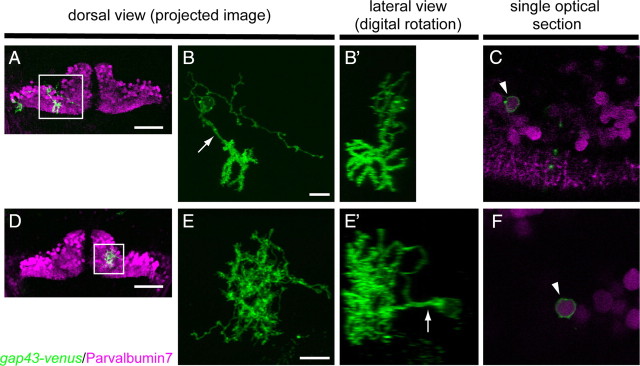
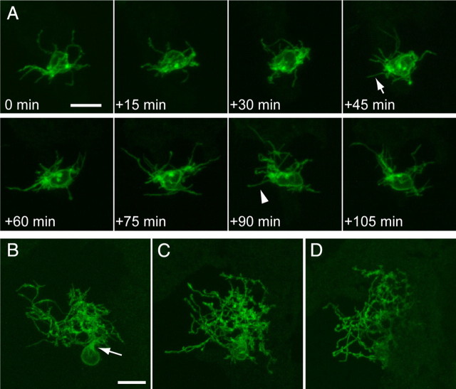
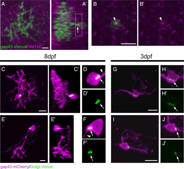
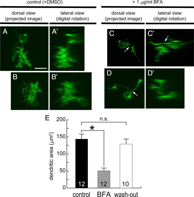
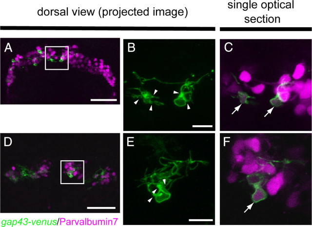
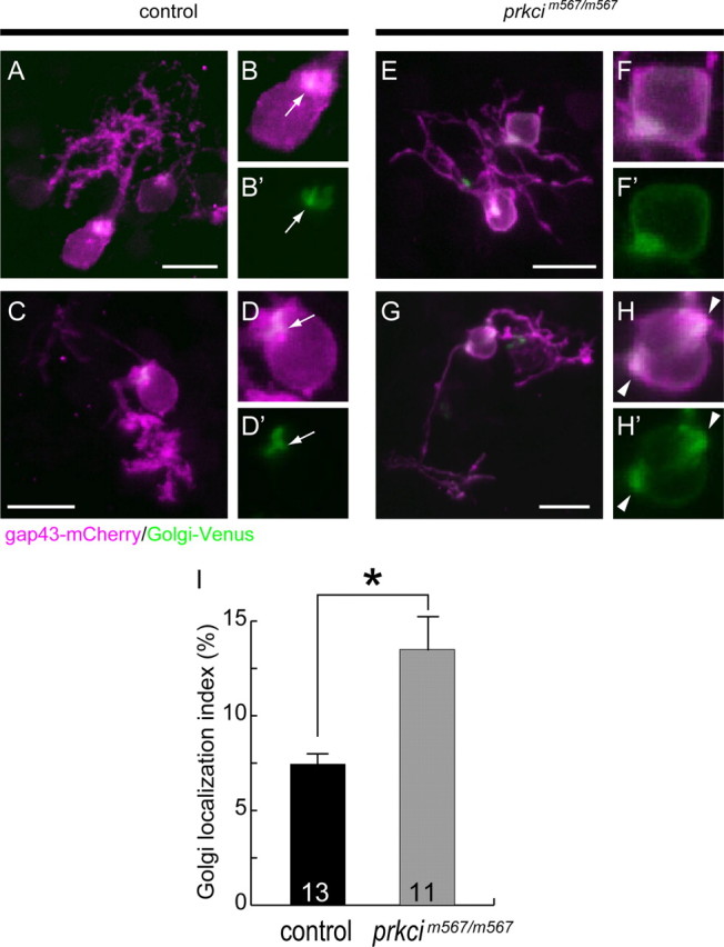
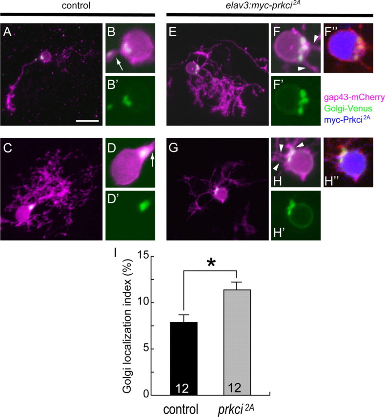
Neurons have highly polarized structures that determine what parts of the soma elaborate the axon and dendrites. However, little is known about the mechanisms that establish neuronal polarity in vivo. Cerebellar Purkinje cells extend a single primary dendrite from the soma that ramifies into a highly branched dendritic arbor. We used the zebrafish cerebellum to investigate the mechanisms by which Purkinje cells acquire these characteristics. To examine dendritic morphogenesis in individual Purkinje cells, we marked the cell membrane using a Purkinje cell-specific promoter to drive membrane-targeted fluorescent proteins. We found that zebrafish Purkinje cells initially extend multiple neurites from the soma and subsequently retract all but one, which becomes the primary dendrite. In addition, the Golgi apparatus specifically locates to the root of the primary dendrite, and its localization is already established in immature Purkinje cells that have multiple neurites. Inhibiting secretory trafficking through the Golgi apparatus reduces dendritic growth, suggesting that the Golgi apparatus is involved in the dendritic morphogenesis. We also demonstrated that in a mutant of an atypical protein kinase C (aPKC), Prkci, Purkinje cells retain multiple primary dendrites and show disrupted localization of the Golgi apparatus. Furthermore, a mosaic inhibition of Prkci in Purkinje cells recapitulates the aPKC mutant phenotype. These results suggest that the aPKC cell autonomously controls the Golgi localization and thereby regulates the specification of the primary dendrite of Purkinje cells.


Figures

Figure 1.
Figure 1.
Dendritic morphology of zebrafish Purkinje cells. A, D, Dorsal view images of the cerebellum of 8 dpf larvae that were injected with aldoca:gap43-Venus plasmid and mosaically expressed gap43-Venus in the Purkinje cells. Two Purkinje cells in A and one in D expressed gap43-Venus (green). All the Purkinje cells were labeled with an anti-parvalbumin7 antibody (magenta). Purkinje cells surrounded by boxes were subjected to observation at a higher resolution (B, B′, C, E, E′, F). Scale bars, 20 μm. B, B′, Dendritic morphology of a relatively posterolateral Purkinje cell labeled with gap43-Venus, boxed in A. B, Projected image of serial confocal optical sections taken from the dorsal side. The Purkinje cell extended a single primary dendrite (arrow) posteriorly. Dense dendritic spines were observed at the dendritic terminals. B′, Lateral view constructed digitally. Dendritic arbors expanded along the dorsoventral axis. Scale bar, 10 μm. E, E′, Dendritic morphology of an anteromedial Purkinje cell, boxed in D. E, Dorsal view with a projected image. E′, Lateral view. The Purkinje cell extended a single primary dendrite dorsally, and the dendritic arbors were randomly distributed. C, F, Single optical sections at the level of the soma of the posterolateral (C) and anteromedial (F) Purkinje cells shown in B and E, respectively. The gap43-Venus-expressing Purkinje cells were positive for parvalbumin7 (magenta). Scale bar, 10 μm.
Figure 2.
Figure 2.
Purkinje cells initially extend multiple neurites and become morphologically mature by retracting these transient neurites. A, Dynamic morphological remodeling of Purkinje cells ∼3 dpf. A medially located Purkinje cell expressing gap43-Venus was subjected to time-lapse confocal imaging. The imaging started at 76 hpf (0 min), and images were taken at 15 min intervals. The neurite indicated by an arrow was newly formed within 15 min, and the one indicated by an arrowhead was retracted within 15 min. B–D, Morphology of the Purkinje cell shown in A at 4, 5, and 6 dpf, respectively. The Purkinje cell completed its primary dendrite specification and extended a single primary dendrite by 4 dpf (B, arrow). After 4 dpf, further dendritic branching took place, and dendritic spines formed. Scale bars: C, D, 10 μm. B–D are shown in the same scale.
Figure 3.
Figure 3.
Golgi apparatus is exclusively localized to the root of the primary dendrite, and this localization precedes the primary dendrite specification. AB′, A Purkinje cell expressing gap43-Venus (green) in an 8 dpf larva that was immunostained with the GM130 antibody (magenta). A, A′, A dorsal view projected image (A) and a lateral view constructed digitally (A′). The GM130-positive Golgi was localized to the root of the primary dendrite (small arrow). B, B′, Higher-magnification views of the projected image of optical sections corresponding to the boxed area in A′. The GM130 signal was only observed at the root of the primary dendrite (arrowheads). C–J′, Purkinje cells in larvae expressing the aldoca:gap43-mCherry-PTV1–2A-Golgi-Venus plasmid. Using the 2A peptide system, the cellular morphology was visualized with gap43-mCherry (magenta), and the Golgi apparatus was visualized with Golgi-Venus (green). C–E′, Purkinje cells in 8 dpf larvae. Dorsal (C, E, projected images) and lateral (C′, E′, constructed digitally) views of anteromedial (C, C′) and posterolateral (E, E′) Purkinje cells. D, D′, and F, F′ are high-magnification views of a single optical section of the Purkinje cells in C and E, respectively. The Golgi apparatuses that were visualized with Golgi-Venus (arrows) were exclusively localized to the root of the primary dendrites (arrowheads). G, I, Dorsal view projected images of Purkinje cells in 3 dpf larvae. H, H′ and J, J′ are high-magnification views of a single optical section in the Purkinje cells in G and I, respectively. At this stage, Purkinje cells had multiple primary dendrites and did not exhibit morphological polarity, but the Golgi apparatus was already localized to a restricted area (arrows). Scale bars, 10 μm.
Figure 4.
Figure 4.
Dendritic growth is suppressed by BFA treatment. A–D′, Purkinje cells expressing gap43-Venus in 5 dpf larvae were treated with DMSO (as control) (A–B′) or 1 μg/ml BFA (C–D′) from 2.5 to 5 dpf. A–D, Dorsal view projected images. A′–D′, Digitally synthesized lateral view images of A–D, respectively. A–B′, A posterolateral Purkinje cell (A, A′) and an anteromedial Purkinje cell (B, B′). Although extensive branching and dense dendritic spines were observed in control Purkinje cells (A, B), the Purkinje cells in BFA-treated larvae had a single primary dendrite (arrows) but exhibited a simple dendritic morphology (C, D). E, Graph of the dendritic area of Purkinje cells in control and BFA-treated larvae. Larvae were treated with DMSO (control, black bar), or 1 μg/ml BFA from 2.5 to 5 dpf (gray bar) or from 2.5 to 4 dpf (BFA was washed out at 4 dpf, white bar). Dendritic area was determined at 5 dpf. Dendritic area was significantly reduced in the Purkinje cells of the larvae treated with BFA from 2.5 to 5 dpf (*p < 0.001 by Welch's t test). Purkinje cells in the larvae that were treated from 2.5 to 4 dpf had a similar size of dendritic area, compared with control Purkinje cells (p > 0.1). Error bars show SEM numbers inside the bars represent the number of cells tested. All images were taken at the same scale. Scale bar, 10 μm.
Figure 5.
Figure 5.
Purkinje cells retain multiple primary dendrites in a prkci mutant. A, D, Dorsal views of 4.5 dpf prkci mutant larvae expressing aldoca:gap43-Venus (green). All the Purkinje cells were stained with an anti-parvalbumin7 antibody (magenta). Scale bars, 50 μm. B, C, E, F, Boxed areas (medial domains) in A and D are shown as high-magnification images in B and E, respectively. B, E, Only the gap43-Venus images are shown. C and F are a single optical section of B and E, respectively, also showing the parvalbumin7 signal. Although the Purkinje cells in the prkci mutant expressed parvalbumin7 (arrows in C, F), they retained multiple primary dendrites (B, E, arrowheads). Scale bars, 10 μm.
Figure 6.
Figure 6.
Golgi localization is disrupted in prkci mutant larvae. A–H′, Purkinje cells from aldoca:gap43-mCherry-PTV1-2A-Golgi-Venus-injected 4.5 dpf control (A–D′) and prkci mutant (E–H′) larvae. A, C, E, G, Anteromedial Purkinje cells (A, E) and posterolateral Purkinje cells (C, G). All images are dorsal view projected images. B, B′, D, D′, F, F′, H, H′, High-magnification views near the soma. In the control Purkinje cells, Golgi-Venus was exclusively localized to the root of a single primary dendrite (B, B′, D, D′, arrows). In contrast, Golgi-Venus was more uniformly distributed in the prkci mutant Purkinje cells, although a strong accumulation was still detected. In one particular case, multiple spots of strong Golgi-Venus accumulation were observed, and two primary dendrites extended from sites adjacent to these accumulations (H, H′, arrowheads). I, Graph of the Golgi localization index of control (black bar) and prkci mutant (gray bar) Purkinje cells. The Golgi localization index was calculated as the percentage of the area of Golgi apparatus over the somal area (see Materials and Methods). The Golgi localization index was significantly increased in the Purkinje cells of prkci mutant larvae (*p < 0.01 by Welch's t test). Error bars show SEM. Numbers inside the bars represent the number of cells tested. Scale bars, 10 μm.
Figure 7.
Figure 7.
Prkci is required cell autonomously for establishing the proper Golgi localization and selection of the primary dendrite. A–H″, Purkinje cells from 5 dpf larvae expressing aldoca:gap43-mCherry-PTV1–2A-Golgi-Venus alone (A–D′, control) or with elavl3:myc-prkci2A (E–H″). All are dorsal view projected images. B, B′, D, D′, F, F′, F″, H, H′, H″, High-magnification views near the soma. Control Purkinje cells extended a single primary dendrite, and Golgi-Venus (green) was exclusively localized to the root of a single primary dendrite (arrows). Purkinje cells that expressed myc-Prkci2A (blue) extended multiple primary dendrites, and the Golgi-Venus+ region was expanded. Extra primary dendrites were extended from adjacent sites to the expanded Golgi (arrowheads). I, Graph of the Golgi localization index of control Purkinje cells (black bar) and Purkinje cells expressing myc-Prkci2A (gray bar). The Golgi localization index was significantly increased in the Purkinje cells expressing myc-Prkci2A (*p < 0.01 in Welch's t test). Error bars show SEM. Numbers inside the bars represent the number of cells tested. All cells are shown in the same scale. Scale bars, 10 μm.

Similar articles

Cited by

References

    1. Altman J, Anderson WJ. Experimental reorganization of the cerebellar cortex. I. Morphological effects of elimination of all microneurons with prolonged x-irradiation started at birth. J Comp Neurol. 1972;146:355–406. - PubMed
    1. Arimura N, Kaibuchi K. Neuronal polarity: from extracellular signals to intracellular mechanisms. Nat Rev Neurosci. 2007;8:194–205. - PubMed
    1. Armengol JA, Sotelo C. Early dendritic development of Purkinje cells in the rat cerebellum. A light and electron microscopic study using axonal tracing in “in vitro” slices. Brain Res Dev Brain Res. 1991;64:95–114. - PubMed
    1. Bae YK, Kani S, Shimizu T, Tanabe K, Nojima H, Kimura Y, Higashijima S, Hibi M. Anatomy of zebrafish cerebellum and screen for mutations affecting its development. Dev Biol. 2009;330:406–426. - PubMed
    1. Baptista CA, Hatten ME, Blazeski R, Mason CA. Cell-cell interactions influence survival and differentiation of purified Purkinje cells in vitro. Neuron. 1994;12:243–260. - PubMed

Publication types

LinkOut - more resources