. 2010 Dec 15;30(50):17004-14.
doi: 10.1523/JNEUROSCI.3513-10.2010.

Golgi cells operate as state-specific temporal filters at the input stage of the cerebellar cortex

Affiliations

Golgi cells operate as state-specific temporal filters at the input stage of the cerebellar cortex

Shane A Heine et al. J Neurosci. .

Abstract

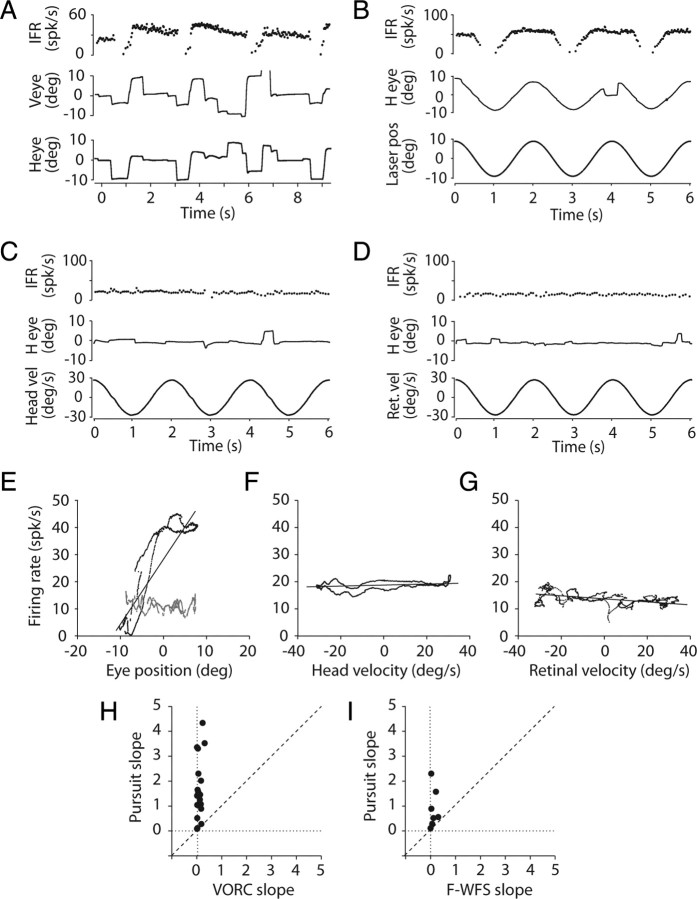
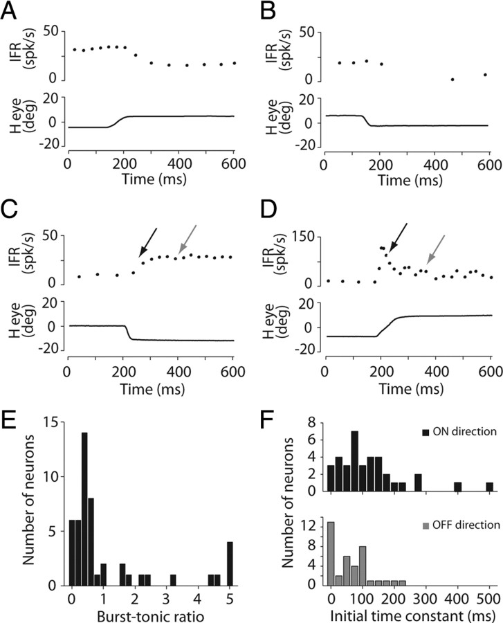
Cerebellar processing of incoming information begins at the synapse between mossy fibers and granule cells, a synapse that is strongly controlled through Golgi cell inhibition. Thus, Golgi cells are uniquely positioned to control the flow of information into the cerebellar cortex and understanding their responses during behavior is essential to understanding cerebellar function. Here we show, for the first time, that Golgi cells express a unique oculomotor-related signal that can be used to provide state- and time-specific filtering of granule cell activity. We used newly established criteria to identify the unique electrophysiological signature of Golgi cells and recorded these neurons in the squirrel monkey ventral paraflocculus during oculomotor behaviors. We found that they carry eye movement, but not vestibular or visual, information and that this eye movement information is only expressed within a specific range of eye positions for each neuron. In addition, simultaneous recordings of Golgi cells and nearby mossy fibers revealed that Golgi cells have the opposite directional tuning of the mossy fiber(s) that likely drive their responses, and that these responses are more sluggish than their mossy fiber counterparts. Because the mossy fiber inputs appear to convey the activity of burst-tonic neurons in the brainstem, Golgi cell responses reflect a time-filtered negative image of the motor command sent to the extraocular muscles. We suggest a role for Golgi cells in the construction of forward models of movement, commonly hypothesized as a major function of the cerebellar cortex in motor control.


Figures

Figure 1.
Figure 1.
Influence of Golgi cells on cerebellar cortex input layer processing and identification of Golgi cells. A, Schematic diagrams illustrating the position of Golgi cells within the cerebellar cortex circuit. Golgi cells receive mossy fiber inputs via their descending dendrites and soma, and parallel fiber inputs via their ascending dendrites. They strongly inhibit granule cells, which are the main glutamatergic input to Purkinje cells. Because granule cells receive inputs from mossy fibers and in turn provide an input to Golgi cells, this circuit configuration gives Golgi cells both feedforward and feedback control over granule cells. B, Location of electrolytic lesion (arrow) placed after recording a putative Golgi cell in the VPFL. The lesion is located in the granular layer (GL), identified as the dark regions in this nissl stain. The Purkinje cell layer (PCL) and molecular layer (ML) are also indicated for reference. Stimulation parameters: 15 uA cathodal current for 15 s. The location of the Golgi cell recordings was confirmed in two additional lesions. C, Scatter plot of median CV2 and median firing rate for 69 Golgi cells (black) and 42 Purkinje cells (gray) identified based on the presence of complex spikes. The corresponding normalized density histograms are shown on the upper and right edges of the axes. spk/s, Spikes/s. D, Histograms of median interspike intervals for all of the Golgi and Purkinje cells (P-cell) shown in C and corresponding spike waveforms for a subset of 10 representative neurons from each group. E, Distribution of Golgi cell distances from Purkinje cell layer for 21 neurons for which adequate depth measurements were taken. See also supplemental Figure S1, available at www.jneurosci.org as supplemental material.
Figure 2.
Figure 2.
Eye movement-only coding by Golgi cells. A, Response during spontaneous eye movements for a representative Golgi cell. Traces, from top, show IFR, vertical eye position (V eye), and horizontal eye position (H eye). B–D, Response of the same neuron during smooth pursuit (B), VOR suppression (C), and F-WFS (D). E–G, Plots of average firing rate versus eye position during pursuit (E), head velocity (vel) during VOR suppression (F), and retinal slip velocity (Ret. vel) during F-WFS with corresponding regression fits (black). E, Average firing rate during horizontal (black) and vertical (gray) pursuit. H, I, slope of regression line during pursuit plotted versus slope of regression line during VOR suppression (VORC; H) or F-WFS (I) for all neurons recorded during these tasks (H, n = 23; I, n = 7). Laser pos, Laser position; spk, spike. See also supplemental Figure S2, available at www.jneurosci.org as supplemental material.
Figure 3.
Figure 3.
Temporal properties of Golgi cell responses. A–D, Representative off (A, B) and on (C, D) responses of four different Golgi cells during saccades. Top, IFR; bottom, horizontal eye position (Heye). E, Distribution of burst–tonic ratios for 49 Golgi cells with significant on responses. Arrows in C and D indicate regions used to calculate burst–tonic ratios (see text). F, Distributions of on (top) and off (bottom) initial time constants for 34 Golgi cells. spk, Spike.
Figure 4.
Figure 4.
Directional tuning of Golgi cells. A, PSTHs of a Golgi cell response to spontaneous saccades within ±45° of each cardinal direction. Gray dotted lines indicate 2 SDs above and below the control firing rate, which was used to calculate the first significant increase or decrease in firing rate, respectively. Bin size is 5 ms. Center plot indicates absolute depth of modulation for each of the four directions. Ipsi, Ipsilateral; contra, contralateral. The distance from the center of the circle to the perimeter equals 20 spikes/s (spk/s). B, C, Number of directions with on (B) or off (C) responses. Each line in the top panel represents a single neuron and the dots indicate the normalized change in firing rate (ΔFR) for saccades in a particular cardinal direction zone. The directions were ranked by response amplitude such that the numbers along the abscissa indicate the most- to least-responsive directions, with direction 1 being the preferred direction. For both B and C, the bottom panel shows the mean and SD for all neurons. See also supplemental Figure S3, available at www.jneurosci.org as supplemental material.
Figure 5.
Figure 5.
Eye position fields of a single Golgi cell. A, Response of Golgi cell during pursuit of a target to the left (left) or right (right) of the center of gaze. Top, IFR; bottom, horizontal eye position (H eye). B, Response of same Golgi cell during spontaneous eye movements. Arrows indicate on response (black) or absence of on response (gray) for two saccades of similar amplitude, but with different starting positions. C, Calculated on (left) and off (right) eye position fields for the same neuron. For both panels, the gray curve indicates changes in firing rate during saccades versus saccade start points and the black curve indicates changes in firing rate during saccades versus saccade end points. The shaded region is the intersection of these two curves, which defines the eye position field of the neuron (see Materials and Methods).
Figure 6.
Figure 6.
Eye-position fields for the population of Golgi cells. A, B, Extent of eye-position response fields for 19 Golgi cells in the on direction (A) and 20 Golgi cells in the off direction (B). Circles indicate borders of position fields. Open circles correspond to estimates of borders that were not clear due to an insufficient number of saccades beyond that position. Gray, Vertical eye-movement cells; black, horizontal eye-movement cells. Positive numbers are up and ipsilateral. Top histograms indicate distributions of eye-position field extents using 1° bins and summing the bins across all neurons. C, Relationship between eye-position response field sizes for on and off directions in 17 Golgi cells. Diagonal line indicates equal sizes. D, Relationship between the eye position at which a Golgi cell first starts to respond in the off direction (upper off border) and stops responding in the on direction (upper on border) for the same 17 Golgi cells. Points falling along the diagonal line indicate that the on and off upper eye position field borders are the same.
Figure 7.
Figure 7.
Relationship between mossy fiber (mf) and Golgi cells (gc) simultaneously recorded during spontaneous eye movements. A, Raw trace from extracellular recording of a mossy fiber–Golgi cell pair on the same electrode (top) and corresponding IFR for the mossy fiber (middle) and Golgi cell (bottom). An upward saccade occurred ∼90 ms and a downward saccade occurred ∼300 ms, producing a burst and then a pause in the mossy fiber firing rate. Note that the Golgi cell appears to be negatively coupled to the mossy fiber. B, This negative coupling is explored further in PSTHs of the same Golgi cell triggered on the mossy fiber burst (top) or pause (bottom). C, The negative coupling between the same mossy fiber–Golgi cell pair is also expressed as opposite directional preferences for on responses (left). This is true for the entire population of nine pairs (right). D, Relationship between Golgi cell off response latencies and mossy fiber on latencies (left), and Golgi cell on response latencies and mossy fiber off latencies (right) for all nine mossy fiber–Golgi cell pairs. Mossy fiber single units are shown as black dots and multiunit hashing is shown as gray dots. Dots falling above the diagonal line indicate that the mossy fiber responds before the Golgi cell. The cluster of three dots (D, right, top) correspond to Golgi cells with on latencies that fall outside the range displayed in the plot. These latencies are 114, 113, and 157 ms. spk, Spike; contra, contralateral; ipsi, ipsilateral.
Figure 8.
Figure 8.
Plausible mechanisms to explain Golgi cell responses. A, Left, Typical mossy fiber burst–tonic response (top) to a change in eye position (pos) resulting from a saccade (middle) and hypothetical firing rate versus eye position curve (bottom). Vertical dashed line indicates mossy-fiber eye-position activation threshold. Right, Typical Golgi cell response for the same eye movement. Note that Golgi cell off response corresponds to mossy fiber burst and on response corresponds to mossy fiber pause. This antiphasic coupling results in the Golgi cell having an inverted eye position response range compared with the mossy fiber (dashed line). B, C, Two possible mechanisms to explain antiphasic coupling of mossy fiber and Golgi cell responses based on known connections and synaptic properties (see text). B, Mechanism 1, Indirect mossy fiber effect over Golgi cell via inhibitory interneurons receiving similarly tuned mossy fiber-granule cell input as Golgi cell. Glutamate (Glu) released from mossy fiber terminals activates ionotropic glutamate receptors (GluR) on Golgi cell and inhibitory interneuron, such as stellate cell. Stellate cell then releases inhibitory neurotransmitter, such as GABA, to generate a Golgi cell firing rate pause in response to mossy fiber burst. GluR activation on the Golgi cell generates initial burst (cf. Fig. 4A) preceding the pause. C, Mechanism 2, Direct mossy fiber effect over Golgi cell via mGluR2 activation of GIRK channels. Glutamate released from mossy fiber terminals activates ionotropic and metabotropic glutamate (i.e., mGluR2) receptors on Golgi cell. The balance between inward current through GluR and outward current through GIRK determines net response of Golgi cell.

Similar articles

Cited by

References

    1. Barmack NH, Yakhnitsa V. Functions of interneurons in mouse cerebellum. J Neurosci. 2008;28:1140–1152. - PMC - PubMed
    1. Blazquez PM, Hirata Y, Heiney SA, Green AM, Highstein SM. Cerebellar signatures of vestibulo-ocular reflex motor learning. J Neurosci. 2003;23:9742–9751. - PMC - PubMed
    1. Chadderton P, Margrie TW, Häusser M. Integration of quanta in cerebellar granule cells during sensory processing. Nature. 2004;428:856–860. - PubMed
    1. Chan-Palay V, Palay SL. The synapse en marron between golgi II neurons and mossy fibers in the rat's cerebellar cortex. Z Anat Entwicklungsgesch. 1971;133:274–287. - PubMed
    1. D'Angelo E, De Zeeuw CI. Timing and plasticity in the cerebellum: focus on the granular layer. Trends Neurosci. 2009;32:30–40. - PubMed

Publication types

LinkOut - more resources