

Mapping brain networks in awake mice using combined optical neural control and fMRI
- PMID: 21160013
- PMCID: PMC3074423
- DOI: 10.1152/jn.00828.2010
Mapping brain networks in awake mice using combined optical neural control and fMRI
Abstract
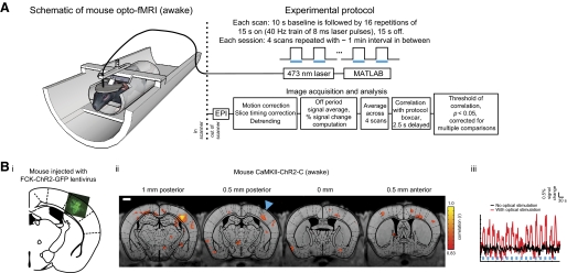
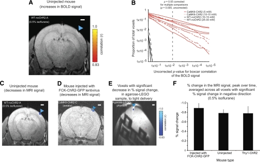
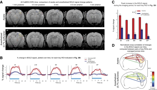
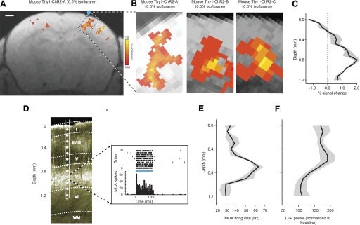
Behaviors and brain disorders involve neural circuits that are widely distributed in the brain. The ability to map the functional connectivity of distributed circuits, and to assess how this connectivity evolves over time, will be facilitated by methods for characterizing the network impact of activating a specific subcircuit, cell type, or projection pathway. We describe here an approach using high-resolution blood oxygenation level-dependent (BOLD) functional MRI (fMRI) of the awake mouse brain-to measure the distributed BOLD response evoked by optical activation of a local, defined cell class expressing the light-gated ion channel channelrhodopsin-2 (ChR2). The utility of this opto-fMRI approach was explored by identifying known cortical and subcortical targets of pyramidal cells of the primary somatosensory cortex (SI) and by analyzing how the set of regions recruited by optogenetically driven SI activity differs between the awake and anesthetized states. Results showed positive BOLD responses in a distributed network that included secondary somatosensory cortex (SII), primary motor cortex (MI), caudoputamen (CP), and contralateral SI (c-SI). Measures in awake compared with anesthetized mice (0.7% isoflurane) showed significantly increased BOLD response in the local region (SI) and indirectly stimulated regions (SII, MI, CP, and c-SI), as well as increased BOLD signal temporal correlations between pairs of regions. These collective results suggest opto-fMRI can provide a controlled means for characterizing the distributed network downstream of a defined cell class in the awake brain. Opto-fMRI may find use in examining causal links between defined circuit elements in diverse behaviors and pathologies.
Figures

References
-
- Aronoff R, Matyas F, Mateo C, Ciron C, Schneider B, Petersen CC. Long-range connectivity of mouse primary somatosensory barrel cortex. Eur J Neurosci 31: 2221–2233, 2010 - PubMed
-
- Ayling O, Harrison T, Boyd J, Goroshkov A, Murphy T. Automated light-based mapping of motor cortex by photoactivation of channelrhodopsin-2 transgenic mice. Nat Methods 6: 219–224, 2009 - PubMed
-
- Bandettini PA, Wong EC, Jesmanowicz A, Hinks RS, Hyde JS. Spin-echo and gradient-echo EPI of human brain activation using BOLD contrast: a comparative study at 1.5 T. NMR Biomed 7: 12–20, 1994 - PubMed
Publication types
MeSH terms
Grants and funding
LinkOut - more resources
Full Text Sources
Other Literature Sources
Medical
Miscellaneous