. 2011 Mar;300(3):C447-55.
doi: 10.1152/ajpcell.00402.2010. Epub 2010 Dec 15.

Fragmented mitochondria are sensitized to Bax insertion and activation during apoptosis

Affiliations

Fragmented mitochondria are sensitized to Bax insertion and activation during apoptosis

Craig Brooks et al. Am J Physiol Cell Physiol. 2011 Mar.

Abstract

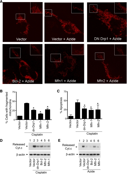
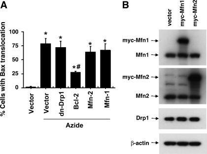
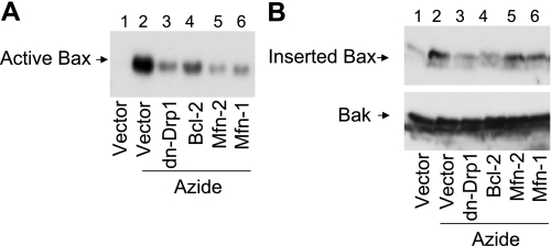
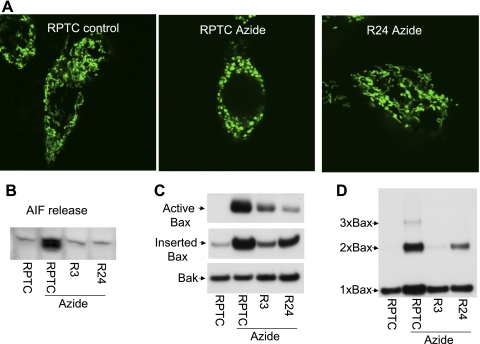
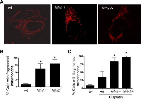
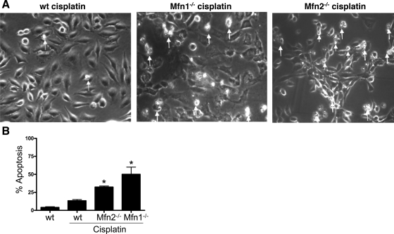
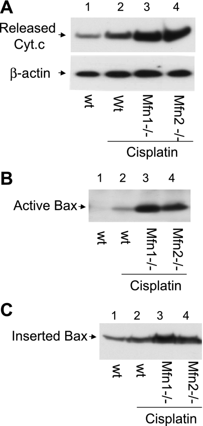
Recent studies have shown mitochondrial fragmentation during cell stress and have suggested a role for the morphological change in mitochondrial injury and ensuing apoptosis. However, the underlying mechanism remains elusive. Here we demonstrate that mitochondrial fragmentation facilitates Bax insertion and activation in mitochondria, resulting in the release of apoptogenic factors. In HeLa cells, overexpression of mitofusins attenuated mitochondrial fragmentation during cisplatin- and azide-induced cell injury, which was accompanied by less apoptosis and less cytochrome c release from mitochondria. Similar effects were shown by inhibiting the mitochondrial fission protein Drp1 with a dominant negative mutant (dn-Drp1). Mitofusins and dn-Drp1 did not seem to significantly affect Bax translocation/accumulation to mitochondria; however, they blocked Bax insertion and activation in mitochondrial membrane. Consistently, in rat kidney proximal tubular cells, small interfering RNA knockdown of Drp1 prevented mitochondrial fragmentation during azide-induced ATP depletion, which was accompanied by less Bax activation, insertion, and oligomerization in mitochondria. These cells released less cytochrome c and AIF from mitochondria and showed significantly lower apoptosis. Finally, mitofusin-null mouse embryonic fibroblasts (MEF) had fragmented mitochondria. These MEFs were more sensitive to cisplatin-induced Bax activation, release of cytochrome c, and apoptosis. Together, this study provides further support for a role of mitochondrial fragmentation in mitochondrial injury and apoptosis. Mechanistically, mitochondrial fragmentation may sensitize the cells to Bax insertion and activation in mitochondria, facilitating the release of apoptogenic factors and consequent apoptosis.


Figures

Fig. 1.
Fig. 1.
Expression of Mfn1, Mfn2, and dn-Drp1 inhibits mitochondrial fragmentation, cytochrome c (Cyt c) release, and apoptosis. A: representative mitochondrial morphology. HeLa cells were cotransfected with Mito-Red and with one of the following plasmids: dn-Drp1, Bcl-2, Mfn1, Mfn2, or empty vector. After overnight transfection, the cells were untreated or treated with 10 mM azide for 3 h. Mitochondrial morphology was examined and recorded by fluorescence microscopy. Insets: boxed area in higher magnification. B: mitochondrial fragmentation during cisplatin treatment. HeLa cells were cotransfected with MitoRed and Mfn1, Mfn2, dn-Drp1, or empty vector. The cells were then treated with 20 μM cisplatin for 16 h to evaluate mitochondrial morphology by fluorescence microscopy. The cells containing fragmented mitochondria and those with filamentous mitochondria were counted to calculate the percentage of cells with mitochondrial fragmentation. C: apoptosis during cisplatin treatment. HeLa cells were transfected as described in A and treated with cisplatin for 24 h to examine apoptosis by morphological criteria. The percentage of apoptosis was determined by counting the cells with typical apoptotic morphology. D: Cyt c release during cisplatin treatment. HeLa cells were transfected as described in A and treated with cisplatin for 24 h. The cells were premeabilized with low concentration digitonin to collect the cytosolic fraction to analyze the released Cyt c by immunoblotting. E: Cyt c release during azide treatment. HeLa cells were transfected as described in A and then subjected to 3 h of ATP depletion with 10 mM azide treatment in glucose-free buffer. The cells were premeabilized with low concentration digitonin to collect the cytosolic fraction to analyze the released Cyt c by immunoblotting. Data in are B and C are expressed as means ± SD (n = 3); *significantly different from the cisplatin-treated empty vector-transfected group.
Fig. 2.
Fig. 2.
DN-Drp1, Mfn1, and Mfn2 do not block Bax translocation to mitochondria during azide treatment. A: Bax translocation analyzed by immunofluorescence staining. HeLa cells were cotransfected with MitoRed and one indicated plasmid (empty vector, dn-Drp-1, Bcl-2, Mfn1, or Mfn2). After azide treatment, the cells were fixed for Bax immunofluorescence staining (labeled by green FITC) and examined to count the cells with Bax translocation to mitochondria. Data are expressed as means ± SD (n = 4); *P < 0.01 vs. control; #P < 0.01 vs. azide treated vector-transfected group. B: Myc-Mfn1 and Myc-Mfn2 expression after transfection. Whole cell lysate was collected for immunoblot analysis using specific antibodies to Mfn1, Mfn2, Drp1, and β-actin.
Fig. 3.
Fig. 3.
DN-Drp1, Mfn1, and Mfn2 suppress Bax activation and insertion in mitochondria membrane. HeLa cells were transfected with Mfn1, Mfn2, dn-Drp1, Bcl-2, or control empty vector. The cells were then subjected to 3 h of 10 mM azide treatment in glucose-free buffer. A: active Bax. Cells lysate was collected for immunoprecipitation with an antibody that was specific to active Bax. The precipitate was finally analyzed by immunoblotting of Bax. B: Bax insertion. Membrane fraction containing mitochondria was collected from the cells and incubated for 30 min with an alkaline (pH 11.5) solution. The mitochondrial fraction was then collected by centrifugation for analysis of remaining Bax by immunoblot analysis. Bak was also analyzed to verify that inserted proteins were not stripped off from mitochondrial membrane by the alkali incubation.
Fig. 4.
Fig. 4.
Knockdown of Drp1 inhibits Bax activation, insertion and oligomerization in mitochondria. R3 and R24 cell clones were generated by stable transfection of Drp-1 short hairpin RNA (shRNA) into rat proximal tubular cells (RPTC). Knockdown of Drp1 in R3 and R24 cells was shown in our recent work. A: representative images of mitochondrial morphology. RPTC and R24 cells were transfected with Mito-Green and then left untreated (control) or treated with 10 mM azide for 3 h to record mitochondrial morphology by fluorescence microscopy. B: apoptosis-inducing factor (AIF) release. Cells were untreated or treated with 10 mM azide for 3 h. The cells were then permeabilized with low concentration digitonin to collect cytosolic fraction for immunoblot analysis of released AIF. C: Bax activation and insertion in mitochondria. RPTC, R3, and R24 cells were untreated or treated with 10 mM azide for 3 h. To analyze Bax activation, cell lysate was collected for immunoprecipitation using an antibody recognizing active Bax, followed by immunoblot analysis of Bax. To analyze Bax insertion, membrane fraction containing mitochondria was isolated and subjected to alkaline (pH 11.5) incubation as described in materials and methods. The fraction was then collected by centrifugation for analysis of remaining Bax by immunoblot analysis. Bak was also analyzed to verify protein loading. D: Bax oligomerization. RPTC, R3, and R24 cells were untreated or treated with 10 mM azide for 3 h. The cells were then cross-linked with 1 mM DSP and further permeabilized with digitonin to collect the membrane fraction containing mitochondria. The membrane fraction was finally subjected to nonreducing gel electrophoresis and immunoblot analysis of Bax.
Fig. 5.
Fig. 5.
Mfn1 or MFn2-null cells have fragmented mitochondria. A: representative mitochondrial morphology. Wild-type (wt), Mfn1-null, and Mfn2-null mouse embryonic fibroblast (MEF) cells were transfected with MitoRed to record mitochondrial morphology by fluorescence microscopy. B: quantification of cells with fragmented mitochondria. Cells were transfected as those cells in A and examined by fluorescence microscopy to determine the percentage of cells with fragmented mitochondria. C: mitochondrial fragmentation during cisplatin treatment. wt, Mfn1-null, and Mfn2-null MEF cells were transfected with MitoRed and then treated with 20 μM cisplatin for 16 h. The cells were examined by fluorescence microscopy to determine the percentage of cells with fragmented mitochondria. Data are means ± SD (n = 3); *significantly different wt cells (B) or cisplatin-treated wt group (C).
Fig. 6.
Fig. 6.
Mfn1 or MF2-null cells are more sensitive to apoptosis. wt, Mfn1-null, and Mfn2-null MEFs were incubated with 20 μM cisplatin for 24 h to record cell morphology. Apoptotic cells were identified by typical morphology including cellular condensation and fragmentation. A: representative cell morphology. Note: many Mfn1 or Mfn2-null cells had undergone apoptosis and detached form the dish. Arrows: representative apoptotic cells. B: percentage of apoptosis. Data are means ± SD (n = 3); *significantly different from wt cells treated with cisplatin.
Fig. 7.
Fig. 7.
Mfn1 or Mfn2-null cells are more sensitive to Cyt c release, Bax activation, and insertion. wt, Mfn1-null, and Mfn2-null MEFs were incubated with 20 μM cisplatin for 24 h. A: Cyt c release. Cells were permeabilized with low concentration digitonin to collect cytosolic fraction for immunoblot analysis to detect Cyt c that had been released into cytosol during cisplatin treatment. B: Bax activation. Cells were lysed with the CHAPS buffer. The lysate was subjected to immunoprecipitation using the antibody specific for active Bax. The resultant immunoprecipitates were analyzed for Bax by immunoblot analysis. C: Bax insertion. Cells were permeabilized with digitonin to release cytosol and collect the membrane fraction with mitochondria, which was subjected to alkaline incubation as described in materials and methods. After alkaline treatment, Bax remaining in the membrane fraction was analyzed by immunoblot analysis.

References

    1. Adams JM, Cory S. Bcl-2-regulated apoptosis: mechanism and therapeutic potential. Current Opin Immunol 19: 488–496, 2007 - PMC - PubMed
    1. Arnoult D, Rismanchi N, Grodet A, Roberts RG, Seeburg DP, Estaquier J, Sheng M, Blackstone C. Bax/Bak-dependent release of DDP/TIMM8a promotes Drp1-mediated mitochondrial fission and mitoptosis during programmed cell death. Curr Biol 15: 2112–2118, 2005 - PubMed
    1. Brooks C, Dong Z. Regulation of mitochondrial morphological dynamics during apoptosis by Bcl-2 family proteins: a key in Bak? Cell Cycle 6: 3043–3047, 2007 - PubMed
    1. Brooks C, Wei Q, Cho S, Dong Z. Regulation of mitochondrial dynamics in acute kidney injury in cell culture and rodent models. J Clin Invest 119: 1275–1285, 2009 - PMC - PubMed
    1. Brooks C, Wei Q, Feng L, Dong G, Tao Y, Mei L, Xie ZJ, Dong Z. Bak regulates mitochondrial morphology and pathology during apoptosis by interacting with mitofusins. Proc Natl Acad Sci USA 104: 11649–11654, 2007 - PMC - PubMed

Publication types

MeSH terms