Comparative Study
. 2010;18(3):173-85.
doi: 10.1159/000322010. Epub 2010 Dec 16.

Long-range regulatory synergy is required to allow control of the TAC1 locus by MEK/ERK signalling in sensory neurones

Affiliations
Comparative Study

Long-range regulatory synergy is required to allow control of the TAC1 locus by MEK/ERK signalling in sensory neurones

Lynne Shanley et al. Neurosignals. 2010.

Abstract

Changes in the expression of the neuropeptide substance P (SP) in different populations of sensory neurones are associated with the progression of chronic inflammatory disease. Thus, understanding the genomic and cellular mechanisms driving the expression of the TAC1 gene, which encodes SP, in sensory neurones is essential to understanding its role in inflammatory disease. We used a novel combination of computational genomics, primary-cell culture and mouse transgenics to determine the genomic and cellular mechanisms that control the expression of TAC1 in sensory neurones. Intriguingly, we demonstrated that the promoter of the TAC1 gene must act in synergy with a remote enhancer, identified using comparative genomics, to respond to MAPK signalling that modulates the expression of TAC1 in sensory neurones. We also reveal that noxious stimulation of sensory neurones triggers this synergy in larger diameter sensory neurones--an expression of SP associated with hyperalgesia. This noxious stimulation of TAC1 enhancer-promotor synergy could be strongly blocked by antagonism of the MEK pathway. This study provides a unique insight into the role of long-range enhancer-promoter synergy and selectivity in the tissue-specific response of promoters to specific signal transduction pathways and suggests a possible new avenue for the development of novel anti-inflammatory therapies.


Figures

Fig. 1
Fig. 1
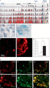
a Plot from the ECR Browser comparing 850 kb surrounding the human TAC1 locus with (from top to bottom) chicken, rat, mouse, dog and rhesus monkey genomes. The VISTA plots represent the genomic extent of (from left to right) the coding regions for ACN9 (homolog of yeast acetate non-utilizing gene 9, involved in gluconeogenesis), TAC1 (tachykinin 1) and ASNS (asparagine synthetase). The xaxis represents linear distance with reference to the human genome sequence. The y-axis represents levels of sequence conservation between 50 and 100%. Blue lines with chevrons represent the genomic extent of each gene. Red, blue, pink and yellow peaks represent areas of sequence conservation (>75% over 100 bp) in intergenic non-coding, exonic, intronic and untranslated regions, respectively (colors in online version only). b, c Whole mount X-galstained DRG preparations from neonate mice transgenic for the ECR2-TAC1prom-LacZ transgene. d, e Florescent immunohistochemical analysis using an anti-SP antibody showing expression in whole mouse neonate DRG neurones after 24 h exposure to vehicle control (d) or 10 μM capsaicin (e). f Bar graph representing the combined results of 3 different experiments on different groups of animals at different times (n = 3) showing proportions of MAP2-expressing cells in DRG neurons that also express SP in the absence (white bar) or presence (black bar) of capsaicin. g i–vi Fluorescence images of an immunohistochemical study of SP and transgene expression on whole DRG explant cultures derived from ECR2-TAC1prom-LacZ transgenic neonates. Cultures represented by i, ii and iii were treated with vehicle and cultures represented by iv, v and vi were treated for 24 h with capsaicin prior to fixing and immunohistochemical analysis. Immunohistochemical analysis was carried out using anti-SP (i and iv) or anti-β-gal (ii and v) as primary antibodies. iii and vi represent merged images where co-localisation is in yellow. White arrows indicate 23 μm.
Fig. 2
Fig. 2
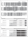
a Sequence alignment of 240 bp of the most highly conserved region of ECR2 highlighting the presence of several conserved transcription factor binding sequences as predicted using the TRANSFAC database. Transcription factor consensus sequences have been highlighted using broken boxes. Sequences conserved back to chicken are highlighted in filled grey boxes. b, c Diagrammatic representation (not to scale) demonstrating the linear relationships of the components of the different luciferase (b) and LacZ constructs (c) used in the current study. pA = SV40 polyadenylation sequence; LacZ = β-galactosidase; hβgprom = human β-globin promoter; TAC1prom = TAC1 promoter; minprom = minimal promoter (Promega).
Fig. 3
Fig. 3
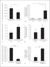
a rtPCR analysis of the levels of TAC1 mRNA derived from whole cultured DRG explants cultured in the presence of absence of the MAPK agonist angiotensin II (n = 3, * p < 0.05). The y-axis represents TAC1 mRNA abundance relative to GAPDH transcripts. b Luciferase reporter gene assay comparing the ability of different promoter constructs described in figure 2b to support luciferase expression in primary sensory neurones. The y-axis represents firefly luciferase activity adjusted to renilla luciferase activity (n = 3, * p < 0.05). c Fold change in the levels of luciferase expression (normalised against renilla luciferase) following angiotensin II treatment of primary DRG neurones transformed with either the TAC1prom-Luc or the ECR2-TAC1prom-Luc constructs (n = 3, * p < 0.05). d Fold change in the number of cells expressing the LacZ marker gene following treatment of primary DRG neurones transformed with either the TAC1prom-LacZ or the ECR2-TAC1prom-LacZ construct and treated with the MAPK agonist angiotensin II (n = 5, * p < 0.05). e, f Dual luciferase reporter studies carried out on primary sensory neurones following their transfection with the ECR2-TAC1prom-Luc construct and normalised using renilla luciferase demonstrating the effects of the MEK antagonist PD98058 (e) and the p38 antagonist SB202190 (f) on the ability of angiotensin II to influence the activity of the ECR2-TAC1prom-Luc (** p < 0.01; *** p < 0.005).
Fig. 4
Fig. 4
a Fold change in the levels of luciferase expression following capsaicin treatment of primary DRG neurones transformed with either the TAC1prom-Luc or the ECR2-TAC1prom-Luc constructs (n = 3, * p < 0.05). b Fold change in the number of cells expressing the LacZ marker gene following capsaicin treatment of primary DRG neurones transformed with either the TAC1prom-LacZ or the ECR2-TAC1prom-LacZ construct (n 1 3, * p < 0.05). c Graphical representation of the proportion of MAP-positive DRG neurones expressing β-gal in ECR2-TAC1prom-LacZ transgenic DRG explants in the absence (white bar) or presence (black bar) of capsaicin (n = 3, * p < 0.05). d Co-localisation of SP expression and ECR2-TAC1prom-LacZ transgene activity in whole transgenic DRG neurones in the presence (white bar) or absence (black bar) of capsaicin treatment (n = 3).
Fig. 5
Fig. 5
a The size distribution (diameter in μm) of neurones within ECR2-TAC1prom-LacZ transgenic neonate DRG explants analysing β-gal expression before and after capsaicin treatment (n = 3) showing a significant shift in the proportion of cells with a diameter greater than 15 μm expressing the transgene. Numbers in brackets represent the total numbers of β-gal expressing cells analysed (n = 3, * p < 0.05). b The proportion of cells of 15 μm in diameter or greater that co-express SP and β-gal following capsaicin treatment (n 1 3). c Dual luciferase reporter studies carried out on primary sensory neurones following their transfection with the ECR2-TAC1prom-Luc construct and normalised using renilla luciferase demonstrating the ability of the MEK antagonist PD98058 to reverse the effects of capsaicin (n = 3, ** p < 0.01, *** p < 0.005).

Similar articles

Cited by

References

    1. Visel A, Rubin EM, Pennacchio LA. Genomic views of distant-acting enhancers. Nature. 2009;461:199–205. - PMC - PubMed
    1. Geppetti P, Nassini R, Materazzi S, Benemei S. The concept of neurogenic inflammation. BJU Int. 2008;101(Suppl 3):2–6. - PubMed
    1. Cao YQ, Mantyh PW, Carlson EJ, Gillespie AM, Epstein CJ, Basbaum AI. Primary afferent tachykinins are required to experience moderate to intense pain. Nature. 1998;392:390–394. - PubMed
    1. Zimmer A, Zimmer AM, Baffi J, Usdin T, Reynolds K, Konig M, Palkovits M, Mezey E. Hypoalgesia in mice with a targeted deletion of the tachykinin 1 gene. Proc Natl Acad Sci USA. 1998;95:2630–2635. - PMC - PubMed
    1. Hokfelt T, Pernow B, Wahren J. Substance p: a pioneer amongst neuropeptides. J Intern Med. 2001;249:27–40. - PubMed

Publication types

MeSH terms