Multicenter Study
. 2011 Mar;79(6):663-670.
doi: 10.1038/ki.2010.484. Epub 2010 Dec 15.

Progressive podocyte injury and globotriaosylceramide (GL-3) accumulation in young patients with Fabry disease

Affiliations
Multicenter Study

Progressive podocyte injury and globotriaosylceramide (GL-3) accumulation in young patients with Fabry disease

Behzad Najafian et al. Kidney Int. 2011 Mar.

Abstract

Progressive renal failure often complicates Fabry disease, the pathogenesis of which is not well understood. To further explore this we applied unbiased stereological quantitative methods to electron microscopic changes of Fabry nephropathy and the relationship between parameters of glomerular structure and renal function in 14 young Fabry patients (median age 12 years). Renal biopsies were obtained shortly before enzyme replacement therapy from these patients and from nine normal living kidney donors as controls. Podocyte globotriaosylceramide (GL-3) inclusion volume density increased progressively with age; however, there were no significant relationships between age and endothelial or mesangial inclusion volume densities. Foot process width, greater in male Fabry patients, also progressively increased with age compared with the controls, and correlated directly with proteinuria. In comparison to the biopsies of the controls, endothelial fenestration was reduced in Fabry patients. Thus, our study found relationships between quantitative parameters of glomerular structure in Fabry nephropathy and age, as well as urinary protein excretion. Hence, podocyte injury may play a pivotal role in the development and progression of Fabry nephropathy.


Figures
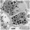
Figure 1
Figure 1
GL-3 inclusions in glomerular podocytes (arrowhead), endothelial cells (double arrowhead) and mesangial cells (spiral arrow) from a kidney biopsy of a Fabry patients (TEM, 11,000 x). Abbreviations: PC=podocyte; C=capillary lumen; E=endothelial cell; M=mesangium; GBM=glomerular basement membrane
Figure 2
Figure 2
Relationship between age and podocyte [Vv(Inc/PC)], mesangial [Vv(Inc/Mes)], and endothelial cell [Vv(Inc/Endo)] GL-3 fractional volume of inclusions per cytoplasm and foot process width (FPW) in Fabry patients. (●=male; ○=female; the bold line is the regression line and the dashed lines are 95% confidence interval boundaries)
Figure 3
Figure 3
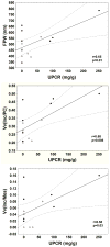
Relationships between urinary protein creatinine ratio (UPCR, mg/g) and foot process width (FPW, nm), GL-3 inclusion fractional volume per podocyte cytoplasm [Vv(Inc/PC)] and mesangial cell cytoplasm [Vv(Inc/Mes)] in Fabry patients. (●=male; ○=female; the bold line is the regression line and the dashed lines are 95% confidence interval boundaries)
Figure 4
Figure 4
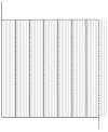
The grid used to estimate % endothelial fenestration composed of an unbiased counting frame with inclusion (dashed) and exclusion (continuous) borders, and parallel lines 4 mm apart. There is one coarse line (where the intercepts with endothelial coverage are counted) per seven fine lines. The short lines on the coarse lines are 4 mm apart and, similar to fine lines, are used to define fenestrated vs. non-fenestrated coverage. The endothelial coverage was arbitrarily called non-fenestrated if the distance between the two fenestrae on either side of the coarse line was more than 3 fine and coarse lines, and otherwise it was called fenestrated.
Figure 5
Figure 5
Comparison of capillary endothelial coverage (arrows) in kidney biopsies from a normal subject (left) and a Fabry patient (right). Note that fenestration is markedly reduced in the Fabry biopsy. Abbreviations: C= capillary; E=endothelial cell; PC=podocyte

Similar articles

Cited by

References

    1. Spada M, Pagliardini S, Yasuda M, et al. High incidence of later-onset Fabry disease revealed by newborn screening. Am J Hum Genet. 2006;79:31–40. - PMC - PubMed
    1. Meikle PJ, Ranieri E, Ravenscroft EM, et al. Newborn screening for lysosomal storage disorders. Southeast Asian J Trop Med Public Health. 1999;30 (Suppl 2):104–110. - PubMed
    1. MacDermot KD, Holmes A, Miners AH. Anderson-Fabry disease: clinical manifestations and impact of disease in a cohort of 98 hemizygous males. J Med Genet. 2001;38:750–760. - PMC - PubMed
    1. MacDermot KD, Holmes A, Miners AH. Anderson-Fabry disease: clinical manifestations and impact of disease in a cohort of 60 obligate carrier females. J Med Genet. 2001;38:769–775. - PMC - PubMed
    1. Wilcox WR, Oliveira JP, Hopkin RJ. Females with Fabry disease frequently have major organ involvement: lessons from the Fabry Registry. Mol Genet Metab. 2008;93:112–28. - PubMed

Publication types

MeSH terms