. 2011 Mar;131(3):706-18.
doi: 10.1038/jid.2010.389. Epub 2010 Dec 16.

Desmosome disassembly in response to pemphigus vulgaris IgG occurs in distinct phases and can be reversed by expression of exogenous Dsg3

Affiliations

Desmosome disassembly in response to pemphigus vulgaris IgG occurs in distinct phases and can be reversed by expression of exogenous Dsg3

Jean M Jennings et al. J Invest Dermatol. 2011 Mar.

Abstract

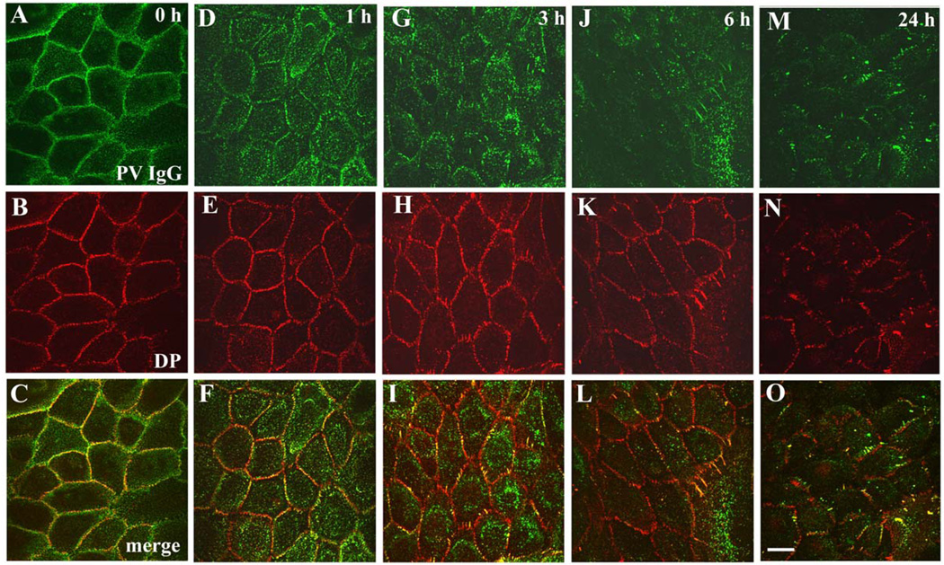
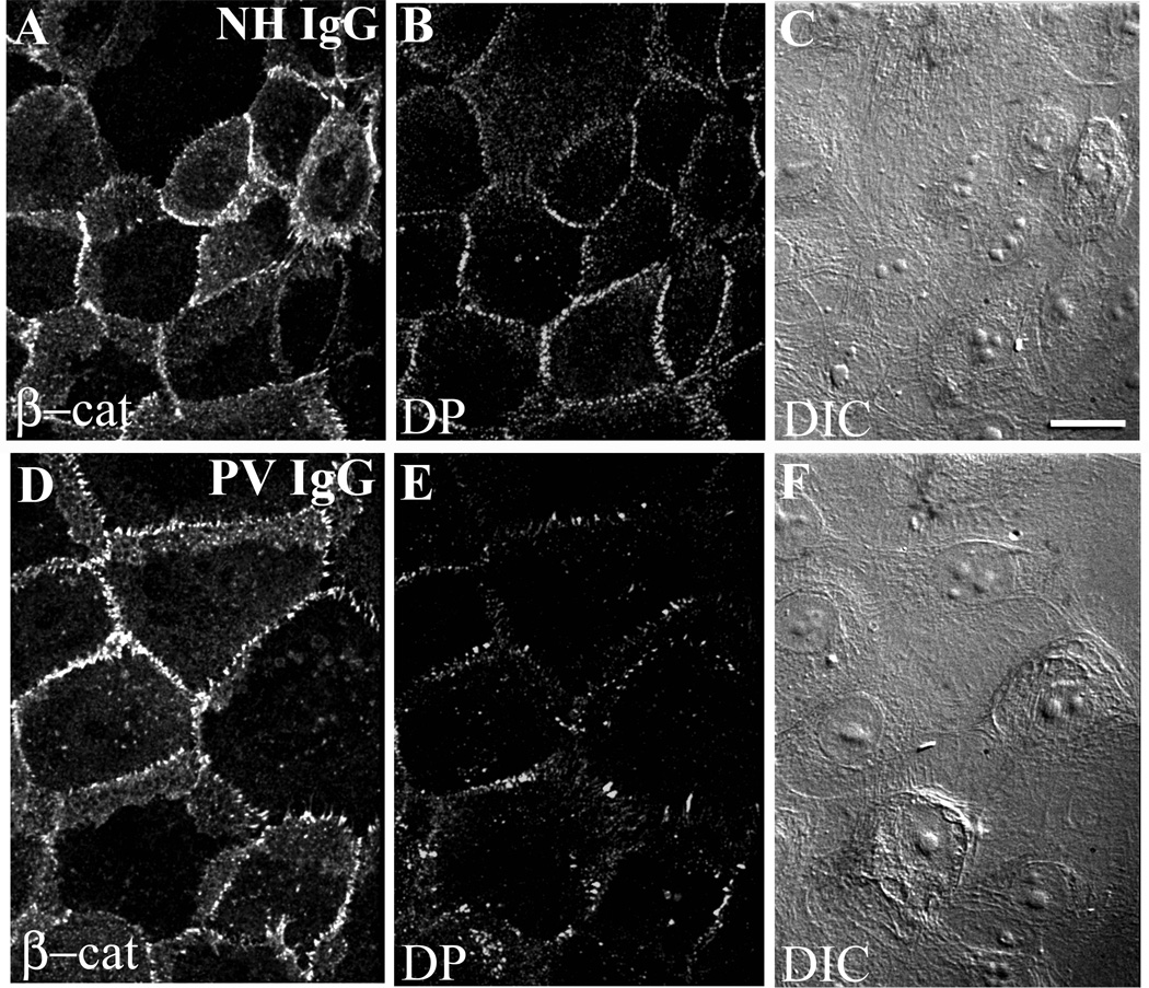
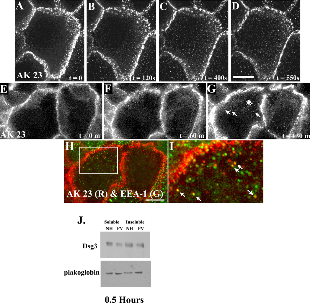
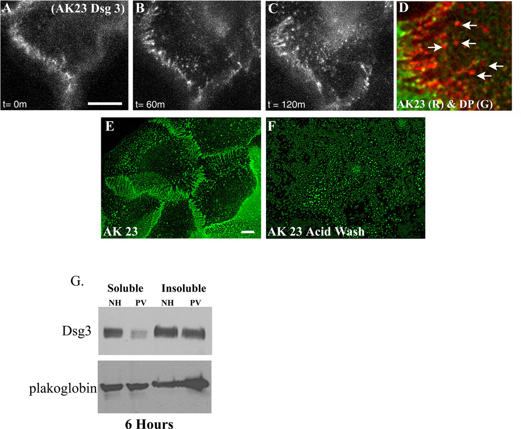
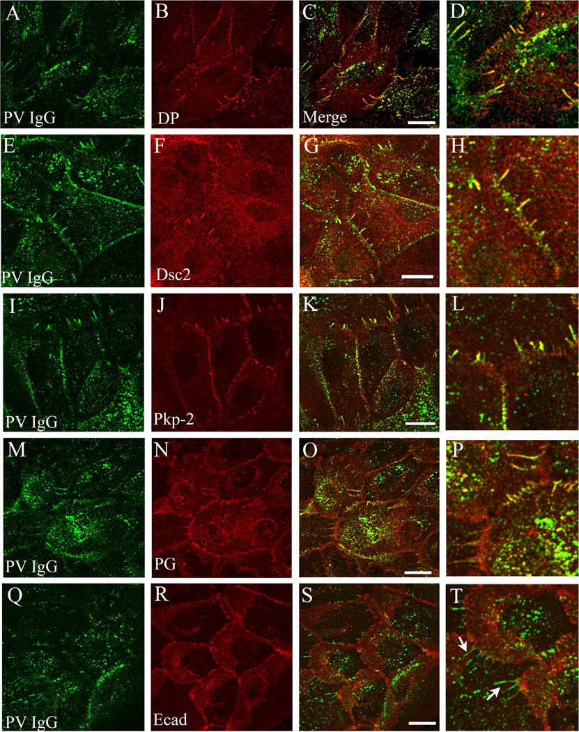
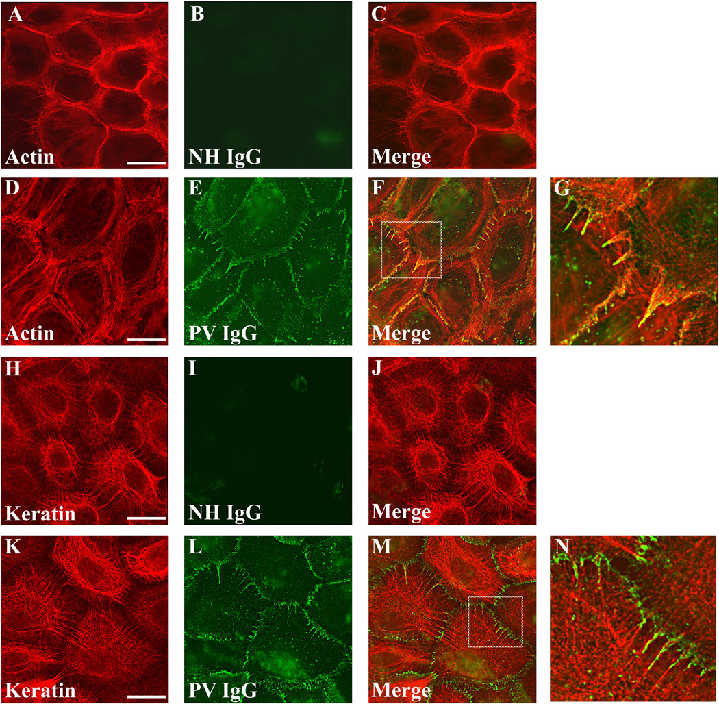
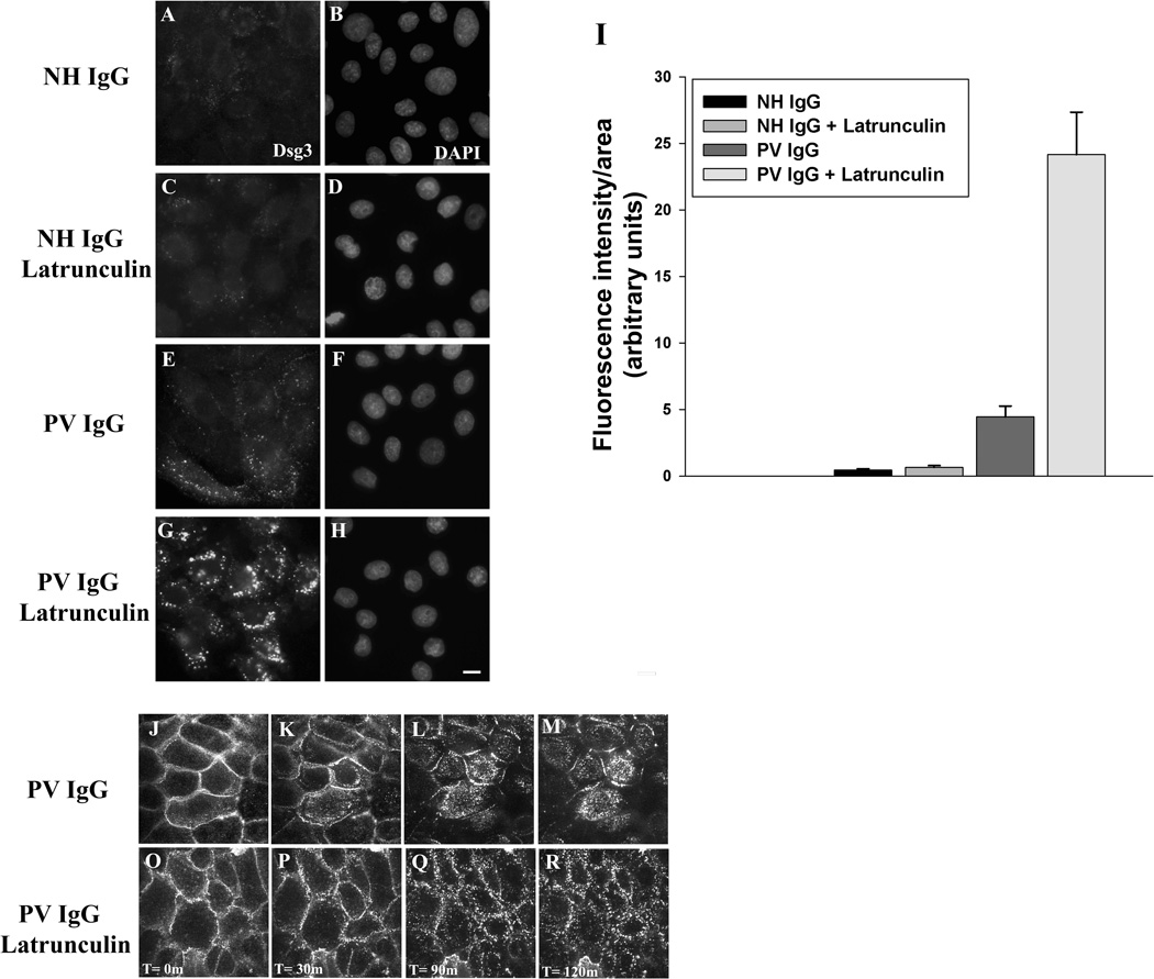
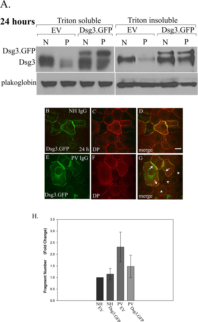
Pemphigus vulgaris (PV) is an epidermal blistering disorder caused by antibodies directed against the desmosomal cadherin desmoglein-3 (Dsg3). The mechanism by which PV IgG disrupts adhesion is not fully understood. To address this issue, primary human keratinocytes (KCs) and patient IgG were used to define the morphological, biochemical, and functional changes triggered by PV IgG. Three phases of desmosome disassembly were distinguished. Analysis of fixed and living KCs demonstrated that PV IgG cause rapid Dsg3 internalization, which likely originates from a non-junctional pool of Dsg3. Subsequently, Dsg3 and other desmosomal components rearrange into linear arrays that run perpendicular to cell contacts. Dsg3 complexes localized at the cell surface are transported in a retrograde manner along with these arrays before being released into cytoplasmic vesicular compartments. These changes in Dsg3 distribution are followed by depletion of detergent-insoluble Dsg3 pools and by the loss of cell adhesion strength. Importantly, this process of disassembly can be prevented by expressing exogenous Dsg3, thereby driving Dsg3 biosynthesis and desmosome assembly. These data support a model in which PV IgG cause the loss of cell adhesion by altering the dynamics of Dsg3 assembly into desmosomes and the turnover of cell surface pools of Dsg3 through endocytic pathways.


Conflict of interest statement

Conflict of Interest: The authors state no conflict of interest.

Figures

Figure 1
Figure 1. Time course of desmosome disassembly in response to PV IgG
Keratinocytes were exposed to PV IgG at 4°C for 20 minutes and subsequently shifted to 37°C for 1, 3, 6, and 24 hours. The localization of human IgG and desmoplakin (DP) was monitored by immunofluorescence microscopy. In cells incubated at 4°C (A–C), PV IgG labels cell borders and desmoplakin staining is predominantly in punctate linear patterns at cell-cell junctions. After 1 hour treatment with PV IgG (D–F), the PV-IgG-Dsg3 molecules accumulate in puncta that are distal to cell-cell borders, while desmoplakin (E) staining is unchanged. Keratinocytes treated with PV IgG for 3 and 6 hours exhibit a rearrangement of desmoplakin into linear arrays emanating from cell borders which contain both Dsg3 and desmoplakin. Following treatment with PV IgG for 24 hours (M–O) both Dsg3 and desmoplakin are noticeably mislocalized and/or absent from cell-cell junctions. Bar, 10µm.
Figure 2
Figure 2. Desmosomes are disrupted by PV IgG but β-catenin is minimally affected and cells remain in close apposition
Keratinocytes were treated with NH IgG (A–C) or PV IgG (D–F) for 6 hours and subsequently processed for immunofluorescence. Note robust β-catenin and desmoplakin (DP) staining (A, B) in cells treated with NH IgG. The PV IgG treated cells exhibit markedly disrupted desmoplakin staining at borders (E), whereas β-catenin remains largely unaltered (D). Differential interference contrast imaging shows that cells do not undergo major changes in cell shape (F). Bar, 10µm.
Figure 3
Figure 3. Non-junctional pools of Dsg3 are rapidly internalized after exposure to PV IgG
The Dsg3 antibody AK23 was fluorescently tagged with Alexa Flour 555 and incubated with human keratinocytes for 30 minutes at 4°C. Unbound antibody was removed and media containing PV IgG was added. Living cells were imaged using time lapse fluorescence microscopy (A–D and E–G) to follow the dynamics of the Dsg3 complex in PV IgG treated cells. Note the appearance of AK23-Dsg3 clusters and vesicular structures that form rapidly after exposure to PV IgG, while junctional Dsg3 remains largely unaltered (Supplemental movie 2). To determine the composition of the AK23-Dsg3 puncta, cells shown in panel G were rapidly fixed and processed for indirect immunofluorescence using antibodies directed against EEA-1. Note co-localization between the AK23-Dsg3 complex and EEA-1 (H, I), indicating that the AK23-Dsg3 complex undergoes internalization to endosomes. Each panel in the series represents a single projection image of 7 (A–D) or 8 (E–G) z-axis images collected at each time point. Sequential detergent extraction and western blot analysis was carried out to determine if membrane associated (Triton X-100 soluble) or cytoskeleton associated (Triton X-100 insoluble) pools of Dsg3 were depleted. A decrease in the soluble pool of Dsg3 was detected within 30 minutes of incubation with PV IgG (J). Results are representative of at least three independently conducted experiments. Bar, 10µm.
Figure 4
Figure 4. The PV IgG-Dsg3 complex reorganizes into linear arrays that exhibit retrograde movement before entering vesicular compartments
Keratinocytes were incubated at 4°C with Alexa Flour labeled AK23 monoclonal antibody for 30 minutes. Unbound antibody was removed followed by the addition of keratinocyte media containing PV IgG. Cells were then imaged every 5 minutes for two hours at 37°C. Notice the formation of linear arrays of AK23-Dsg3, followed by apparent budding of vesicular structures from the tips of the arrays (B, C). (Supplemental movie 3). Retrospective immunofluorescence analysis (D) indicates that Dsg3 that has entered this vesicular compartment is no longer associated with desmoplakin (arrows, panel D). To determine if Dsg3 in the linear arrays was present on the cell surface, living cells were incubated with the pathogenic monoclonal AK23 for 3 hrs in the presence of PV IgG. Cells were then incubated on ice and either fixed immediately (E) or incubated in a low pH acid wash before fixation (F) to remove cell surface bound AK23 IgG. Note that the linear arrays are not observed in cells exposed to the low pH wash, demonstrating that Dsg3 in these structures is present at the cell surface and that the punctate vesicular Dsg3 observed in panel F is intracellular. Western blot analysis of keratinocyte lysates treated for 6 hrs in PV IgG reveals a decrease in the non-junctional Triton X-100 soluble pool of Dsg3, whereas the Triton insoluble pool of Dsg3 exhibited little or no change. No changes in plakoglobin levels were observed (G). Results are representative of at least three independently conducted experiments. Bar, 10µm.
Figure 5
Figure 5. Linear arrays contain desmosomal but not adherens junction components
Primary uman keratinocytes were incubated with PV IgG for 6 hours and then processed for confocal immunofluorescence microscopy for PV IgG and either desmoplakin (DP, A–D), desmocollin-2 (Dsc2, E–H), plakophilin-2 (Pkp-2, I–L), plakoglobin (M–P), or E-cadherin (Ecad, Q–T). Note that the PV IgG complex colocalizes with each of the desmosomal components in linear arrays, but does not colocalize with the adherens junction marker E-cadherin (arrows panel T). Bar, 10µm.
Figure 6
Figure 6. PV IgG-Dsg3 in linear arrays colocalize actin and align with keratin filaments
Keratinocytes were treated with either NH IgG or PV IgG for 6 hrs and processed for immunofluorescence localization of NH and PV IgG with actin (A–G) or NH and PV IgG and cytokeratin (H–N). Note colocalization of PV IgG in linear arrays with actin, which emanate away from cell cell borders in alignment with keratin filaments. Images shown are single z-plane representations that were captured using wide field microscopy followed by deconvolution. Bar, 20µm.
Figure 7
Figure 7. Actin depolymerization increases PV IgG-induced Dsg3 internalization
Keratinocytes were untreated (A–B and E–F) or treated with latrunculin A (250 nM) (C–D and G–H) at 37°C for 1 hour. Cells were then incubated at 4°C for 30 min with AK23 IgG to label cell surface pools of Dsg3. Excess AK23 antibody was removed from the medium, NH IgG or PV IgG were added, and the cells were shifted to 37°C for 3 hours. To visualize only the intracellular pool of Dsg3, cells were washed in a low pH buffer at 4°C to remove surface bound antibody prior to processing for immunofluorescence. Bar, 5µm. The amount of internalized AK23-Dsg3 was then quantified (I). Error bars represent the standard error of the mean, where n = 15 fields of view. In parallel experiments, time lapse microscopy was used to visualize Dsg3 dynamics in PV IgG (J–M) or PV IgG + Latrunculin A (O–R) treated keratinocytes (Supplemental movies 4 and 5).
Figure 8
Figure 8. Expression of exogenous Dsg3.GFP prevents desmoplakin mislocalization and loss of cell adhesion in PV IgG treated keratinocytes
Dsg3.GFP was expressed in keratinocytes using an adenoviral delivery system 24 hours prior to treatment with NH or PV IgG. After an additional 24 hrs, cells were processed for sequential detergent extraction and western blot (Panel A), immunofluorescence (Panels B–G), or subjected to mechanical stress to measure cell adhesion strength (Panel H). Note that expression of exogenous Dsg3.GFP prevented the down-regulation of Dsg3 in both the Triton X-100 soluble and insoluble pools in PV IgG (P) treated cells compared to cells treated with NH IgG (N) (Panel A). Note that Dsg3.GFP colocalized with desmoplakin in NH IgG treated cells (B–D) and prevented mislocalization of desmoplakin in PV IgG treated cells (E–G). Note also the robust desmoplakin staining in cells that are expressing Dsg3.GFP (arrows, G) in sharp contrast to adjacent cells lacking Dsg3.GFP (arrowheads, G). To determine if Dsg3.GFP expression could prevent loss of cell adhesion strength in PV IgG treated cells, NH IgG or PV IgG treated keratinocytes were released from the substrate using the enzyme dispase and subjected to mechanical stress. The number of resulting monolayer fragments was quantified. Note that exogenous Dsg3.GFP rendered PV IgG treated keratinocytes resistant to mechanical stress. The graph represents an average of three separate experiments quantified by a blinded observer. PV IgG treated keratinocytes expressing Dsg3.GFP exhibited significantly less fragments than those expressing empty adenovirus as determined using ANOVA with post hoc least significant difference analysis (**p < 0.01). Error bars represent the standard deviation. Bar, 10µm.

References

    1. Amagai M. Pemphigus vulgaris and its active disease mouse model. Curr Dir Autoimmun. 2008;10:167–181. - PubMed
    1. Awad MM, Calkins H, Judge DP. Mechanisms of disease: molecular genetics of arrhythmogenic right ventricular dysplasia/cardiomyopathy. Nat Clin Pract Cardiovasc Med. 2008;5:258–267. - PMC - PubMed
    1. Bass-Zubek AE, Hobbs RP, Amargo EV, Garcia NJ, Hsieh SN, Chen X, et al. Plakophilin 2: a critical scaffold for PKC alpha that regulates intercellular junction assembly. J Cell Biol. 2008;181:605–613. - PMC - PubMed
    1. Bazzi H, Christiano AM. Broken hearts, woolly hair, and tattered skin: when desmosomal adhesion goes awry. Curr Opin Cell Biol. 2007;19:515–520. - PMC - PubMed
    1. Berkowitz P, Hu P, Liu Z, Diaz LA, Enghild JJ, Chua MP, et al. Desmosome signaling. Inhibition of p38MAPK prevents pemphigus vulgaris IgG-induced cytoskeleton reorganization. J Biol Chem. 2005;280:23778–23784. - PubMed

Publication types

MeSH terms