

Exercise echocardiography
- PMID: 21160588
- PMCID: PMC2998822
- DOI: 10.4330/wjc.v2.i8.223
Exercise echocardiography
Abstract
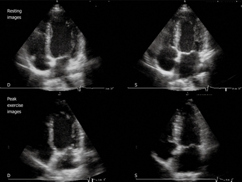
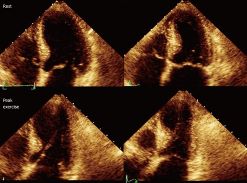
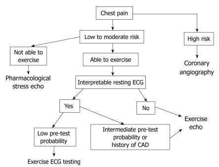
Exercise echocardiography has been used for 30 years. It is now considered a consolidated technique for the diagnosis and risk stratification of patients with known or suspected coronary artery disease (CAD). Of the stress echocardiography techniques, it represents the first choice for patients who are able to exercise. Given that the cost-effectiveness and safety of stress echocardiography are higher than those of other imaging techniques, its use is likely to be expanded further. Recent research has also proposed this technique for the evaluation of cardiac pathology beyond CAD. Although the role of new technology is promising, the assessment of cardiac function relies on good quality black and white harmonic images.
Keywords: Coronary artery disease; Exercise echocardiography; Peak imaging.
Figures
References
-
- Gibbons RJ, Abrams J, Chatterjee K, Daley J, Deedwania PC, Douglas JS, Ferguson TB Jr, Fihn SD, Fraker TD Jr, Gardin JM, et al. ACC/AHA 2002 guideline update for the management of patients with chronic stable angina--summary article: a report of the American College of Cardiology/American Heart Association Task Force on practice guidelines (Committee on the Management of Patients With Chronic Stable Angina) J Am Coll Cardiol. 2003;41:159–168. - PubMed
-
- Pellikka PA, Nagueh SF, Elhendy AA, Kuehl CA, Sawada SG. American Society of Echocardiography recommendations for performance, interpretation, and application of stress echocardiography. J Am Soc Echocardiogr. 2007;20:1021–41. - PubMed
-
- Fox K, Garcia MA, Ardissino D, Buszman P, Camici PG, Crea F, Daly C, De Backer G, Hjemdahl P, Lopez-Sendon J, et al. Guidelines on the management of stable angina pectoris: executive summary: The Task Force on the Management of Stable Angina Pectoris of the European Society of Cardiology. Eur Heart J. 2006;27:1341–1381. - PubMed
-
- Wann LS, Faris JV, Childress RH, Dillon JC, Weyman AE, Feigenbaum H. Exercise cross-sectional echocardiography in ischemic heart disease. Circulation. 1979;60:1300–1308. - PubMed
-
- Burgess MI, Jenkins C, Sharman JE, Marwick TH. Diastolic stress echocardiography: hemodynamic validation and clinical significance of estimation of ventricular filling pressure with exercise. J Am Coll Cardiol. 2006;47:1891–1900. - PubMed
LinkOut - more resources
Full Text Sources
Miscellaneous
