

Clinical use of nuclear cardiology in the assessment of heart failure
- PMID: 21160612
- PMCID: PMC2999043
- DOI: 10.4330/wjc.v2.i10.344
Clinical use of nuclear cardiology in the assessment of heart failure
Abstract
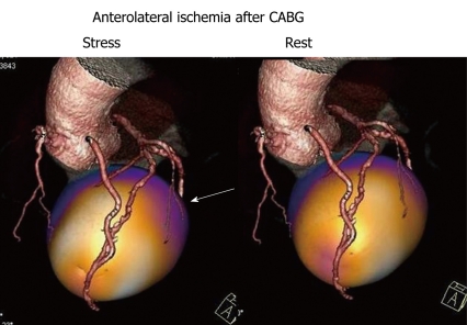
A nuclear cardiology test is the most commonly performed non-invasive cardiac imaging test in patients with heart failure, and it plays a pivotal role in their assessment and management. Quantitative gated single positron emission computed tomography (QGS) is used to assess quantitatively cardiac volume, left ventricular ejection fraction (LVEF), stroke volume, and cardiac diastolic function. Resting and stress myocardial perfusion imaging, with exercise or pharmacologic stress, plays a fundamental role in distinguishing ischemic from non-ischemic etiology of heart failure, and in demonstrating myocardial viability. Diastolic heart failure also termed as heart failure with a preserved LVEF is readily identified by nuclear cardiology techniques and can accurately be estimated by peak filling rate (PFR) and time to PFR. Movement of the left ventricle can also be readily assessed by QGS, with newer techniques such as three-dimensional, wall thickening evaluation aiding its assessment. Myocardial perfusion imaging is also commonly used to identify candidates for implantable cardiac defibrillator and cardiac resynchronization therapies. Neurotransmitter imaging using (123)I-metaiodobenzylguanidine offers prognostic information in patients with heart failure. Metabolism and function in the heart are closely related, and energy substrate metabolism is a potential target of medical therapies to improve cardiac function in patients with heart failure. Cardiac metabolic imaging using (123)I-15-(p-iodophenyl)3-R, S-methylpentadecacoic acid is a commonly used tracer in clinical studies to diagnose metabolic heart failure. Nuclear cardiology tests, including neurotransmitter imaging and metabolic imaging, are now easily preformed with new tracers to refine heart failure diagnosis. Nuclear cardiology studies contribute significantly to guiding management decisions for identifying cardiac risk in patients with heart failure.
Keywords: Diastolic function; Metaiodobenzylguanidine; Prognosis; Quantitative gated single photon emission computed tomography; β-methyl-p-iodophenyl-pentadecanoic acid.
Figures
Similar articles
-
Echocardiography in the assessment of heart failure.Minerva Cardioangiol. 2009 Aug;57(4):457-66. Minerva Cardioangiol. 2009. PMID: 19763068 Review.
-
Clinical usefulness of a novel program "Heart Function View" for evaluating cardiac function from gated myocardial perfusion SPECT.Ann Nucl Med. 2014 Oct;28(8):812-23. doi: 10.1007/s12149-014-0875-0. Epub 2014 Jul 15. Ann Nucl Med. 2014. PMID: 25023232
-
Clinical significance of diastolic function as an indicator of myocardial ischemia assessed by 16-frame gated myocardial perfusion SPECT.Ann Nucl Med. 2008 Oct;22(8):677-83. doi: 10.1007/s12149-008-0174-8. Epub 2008 Nov 4. Ann Nucl Med. 2008. PMID: 18982470
-
Cardiac function, perfusion, and morbidity in irradiated long-term survivors of Hodgkin's disease.Int J Radiat Oncol Biol Phys. 1997 Nov 1;39(4):897-906. doi: 10.1016/s0360-3016(97)00467-7. Int J Radiat Oncol Biol Phys. 1997. PMID: 9369139
-
[Role of perfusion myocardial scintigraphy with gated SPECT technique in the diagnostic and prognostic evaluation of patients with chronic coronary disease].Ital Heart J Suppl. 2002 Mar;3(3):309-18. Ital Heart J Suppl. 2002. PMID: 12040846 Review. Italian.
Cited by
-
A new perspective on an old method: gated SPECT imaging for left ventricular contractile function assessment.J Nucl Cardiol. 2023 Dec;30(6):2658-2665. doi: 10.1007/s12350-023-03340-1. Epub 2023 Jul 25. J Nucl Cardiol. 2023. PMID: 37491510
-
Evaluation of Cardiac Mitochondrial Function by a Nuclear Imaging Technique using Technetium-99m-MIBI Uptake Kinetics.Asia Ocean J Nucl Med Biol. 2013 Spring;1(1):39-43. doi: 10.7508/aojnmb.2013.01.008. Asia Ocean J Nucl Med Biol. 2013. PMID: 27408841 Free PMC article. Review.
-
Imaging in Heart Failure with Preserved Ejection Fraction: A Multimodality Imaging Point of View.Card Fail Rev. 2023 Apr 4;9:e04. doi: 10.15420/cfr.2022.27. eCollection 2023. Card Fail Rev. 2023. PMID: 37387734 Free PMC article. Review.
-
Computed tomography of cardiomyopathies.Cardiovasc Diagn Ther. 2017 Oct;7(5):539-556. doi: 10.21037/cdt.2017.09.07. Cardiovasc Diagn Ther. 2017. PMID: 29255695 Free PMC article. Review.
-
Non-Invasive Assessment of Left Ventricle Ejection Fraction: Where Do We Stand?J Pers Med. 2021 Nov 5;11(11):1153. doi: 10.3390/jpm11111153. J Pers Med. 2021. PMID: 34834505 Free PMC article. Review.
References
-
- Hogg K, Swedberg K, McMurray J. Heart failure with preserved left ventricular systolic function; epidemiology, clinical characteristics, and prognosis. J Am Coll Cardiol. 2004;43:317–327. - PubMed
-
- Owan TE, Hodge DO, Herges RM, Jacobsen SJ, Roger VL, Redfield MM. Trends in prevalence and outcome of heart failure with preserved ejection fraction. N Engl J Med. 2006;355:251–259. - PubMed
-
- Matsuo S, Nakae I, Tsutamoto T, Okamoto N, Horie M. A novel clinical indicator using Tc-99m sestamibi for evaluating cardiac mitochondrial function in patients with cardiomyopathies. J Nucl Cardiol. 2007;14:215–220. - PubMed
-
- Matsuo S, Matsumoto T, Nakae I, Koh T, Masuda D, Takada M, Murata K, Horie M. Prognostic value of ECG-gated thallium-201 single-photon emission tomography in patients with coronary artery disease. Ann Nucl Med. 2004;18:617–622. - PubMed
-
- Germano G, Kavanagh PB, Slomka PJ, Van Kriekinge SD, Pollard G, Berman DS. Quantitation in gated perfusion SPECT imaging: the Cedars-Sinai approach. J Nucl Cardiol. 2007;14:433–454. - PubMed
LinkOut - more resources
Full Text Sources
