

Acute pancreatitis secondary to intramural duodenal hematoma: Case report and literature review
- PMID: 21160669
- PMCID: PMC2998857
- DOI: 10.4329/wjr.v2.i7.283
Acute pancreatitis secondary to intramural duodenal hematoma: Case report and literature review
Abstract
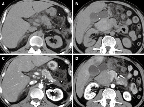
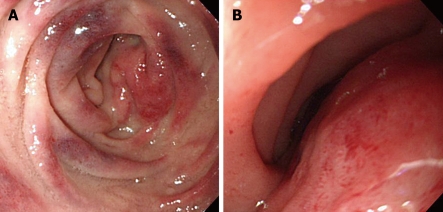
Nontraumatic intramural duodenal hematoma (IDH) is rare disease and it is generally related to coagulation abnormalities. Reports of nontraumatic IDH associated with pancreatic disease are relatively rare, and various conditions including acute or chronic pancreatitis are thought to be associated with nontraumatic IDH. However, the association between IDH and acute pancreatitis remains unknown. We report the case of a 45-year-old man who presented with vomiting and right hypochondrial pain. He had no medical history, but was a heavy drinker. The diagnosis of IDH was established by computed tomography, ultrasonography and endoscopy, and it was complicated by acute pancreatitis. The lesions resolved with conservative management. We discuss this case in the context of previously reported cases of IDH concomitant with acute pancreatitis. In our patient, acute pancreatitis occurred concurrently with hematoma, probably due to obstruction of the duodenal papilla, or compression of the pancreas caused by the hematoma. The present analysis of the published cases of IDH with acute pancreatitis provides some information on the pathogenesis of IDH and its relationship with acute pancreatitis.
Keywords: Acute pancreatitis; Computed tomography; Intramural duodenal hematoma; Jaundice; Ultrasonography.
Figures

References
-
- M’Lauchlan J. False aneurysm tumour occupying nearly the whole of the duodenum. Lancet. 1838;2:203–205.
-
- Hayashi K, Futagawa S, Kozaki S, Hirao K, Hombo Z. Ultrasound and CT diagnosis of intramural duodenal hematoma. Pediatr Radiol. 1988;18:167–168. - PubMed
-
- Jewett TC Jr, Caldarola V, Karp MP, Allen JE, Cooney DR. Intramural hematoma of the duodenum. Arch Surg. 1988;123:54–58. - PubMed
-
- Aston JK. Computed tomography of obstructive jaundice secondary to duodenal hematoma. J Comput Tomogr. 1986;10:171–173. - PubMed
-
- Antoniou D, Zarifi M, Gentimi F, Christopoulos-Geroulanos G. Sonographic diagnosis and monitoring of an intramural duodenal hematoma following upper endoscopic biopsy in a child. J Clin Ultrasound. 2009;37:534–538. - PubMed
LinkOut - more resources
Full Text Sources
