

Actinomycosis of the sigmoid colon: A case report
- PMID: 21160798
- PMCID: PMC2999117
- DOI: 10.4240/wjgs.v1.i1.62
Actinomycosis of the sigmoid colon: A case report
Abstract
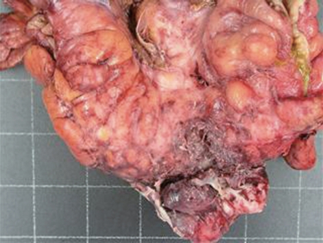
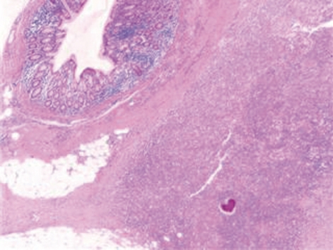
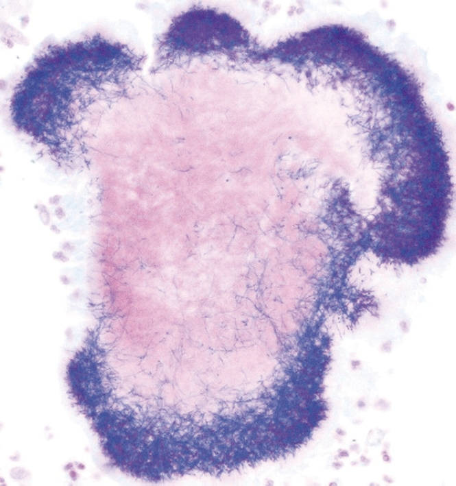
Abdominal actinomycosis is a chronic suppurative infection caused by Actinomyces species. The ileo-cecal region is most commonly affected, while the left side of the colon is more rarely involved. The infection has a tendency to infiltrate adjacent tissues and is therefore rarely confined to a single organ. Presentation may vary from non specific symptoms and signs to an acute abdomen. A computed tomography scan is helpful in identifying the inflammatory process and the organs involved. It also allows visual guidance for percutaneous drainage of abscesses, thus aiding diagnosis. Culture is difficult because of the anaerobic character and slow growth of actinomycetes. Colonoscopy is usually normal, but may shows signs of external compression. Preoperative diagnosis is rare and is established only in less than 10% of cases. In uncomplicated disease, high dose antibiotic therapy is the mainstay of treatment. Surgery is often performed because of a difficulty in diagnosis. Surgery and antibiotics are required in the case of complicated disease. Combined medical and surgical treatment achieves a cure in about 90% of cases. The authors report a case of sigmoid actinomycosis where diagnosis was made from the histology, and a review of the literature is presented.
Keywords: Abdominal pain; Actinomycosis; Gram-positive bacteria; Sigmoid colon; Sulfur.
Figures
References
-
- Brown JR. Human actinomycosis. A study of 181 subjects. Hum Pathol. 1973;4:319–330. - PubMed
-
- Israel J. Neue Beobachtungen auf dem Gebiete der Mykosen des Menschen. Archiv Path Anat Physiol Klin Med. 1978;74:15–53.
-
- Kaya E, Yilmazlar T, Emiroğlu Z, Zorluoğlu A, Bayer A. Colonic actinomycosis: report of a case and review of the literature. Surg Today. 1995;25:923–926. - PubMed
-
- Coremans G, Margaritis V, Van Poppel HP, Christiaens MR, Gruwez J, Geboes K, Wyndaele J, Vanbeckevoort D, Janssens J. Actinomycosis, a rare and unsuspected cause of anal fistulous abscess: report of three cases and review of the literature. Dis Colon Rectum. 2005;48:575–581. - PubMed
-
- de Feiter PW, Soeters PB. Gastrointestinal actinomycosis: an unusual presentation with obstructive uropathy: report of a case and review of the literature. Dis Colon Rectum. 2001;44:1521–1525. - PubMed
LinkOut - more resources
Full Text Sources
