

Jejunal diverticular disease complicated by enteroliths: Report of two different presentations
- PMID: 21160831
- PMCID: PMC2999198
- DOI: 10.4240/wjgs.v2.i1.26
Jejunal diverticular disease complicated by enteroliths: Report of two different presentations
Abstract
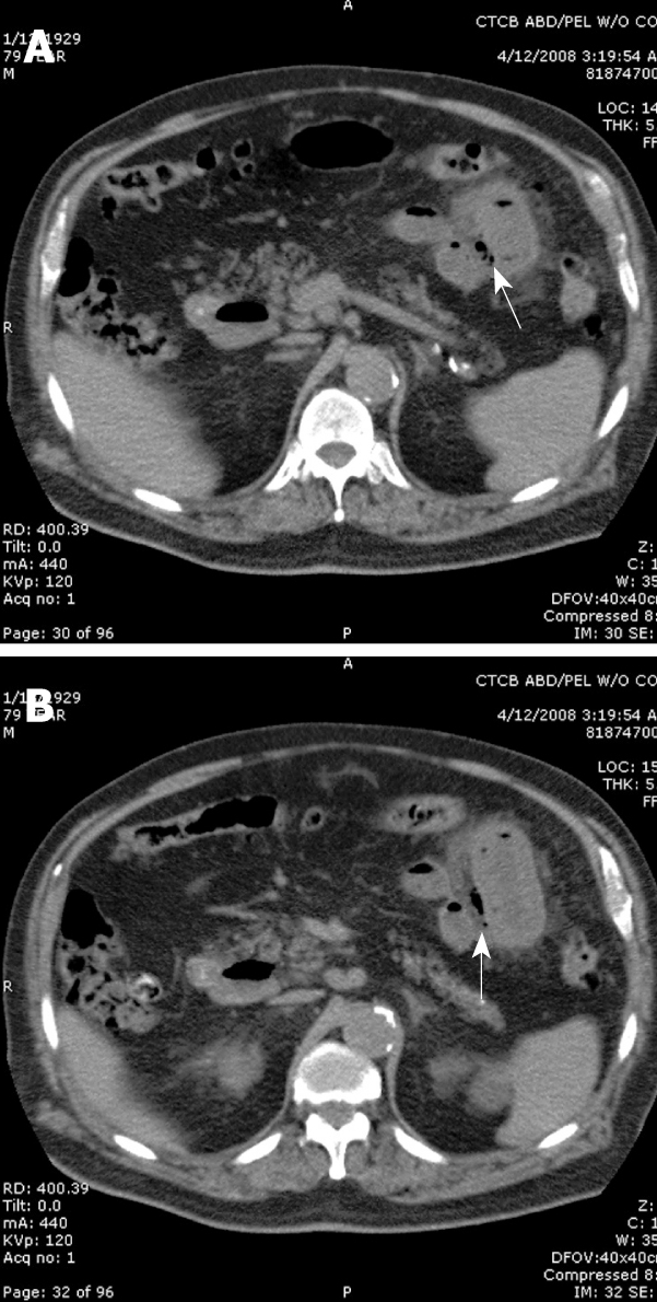
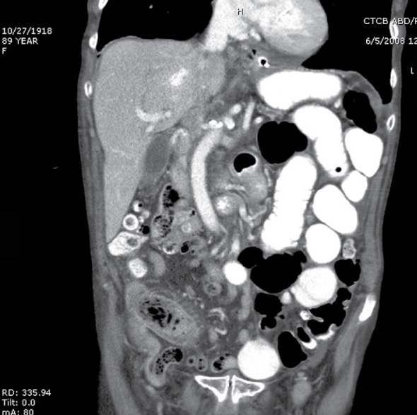
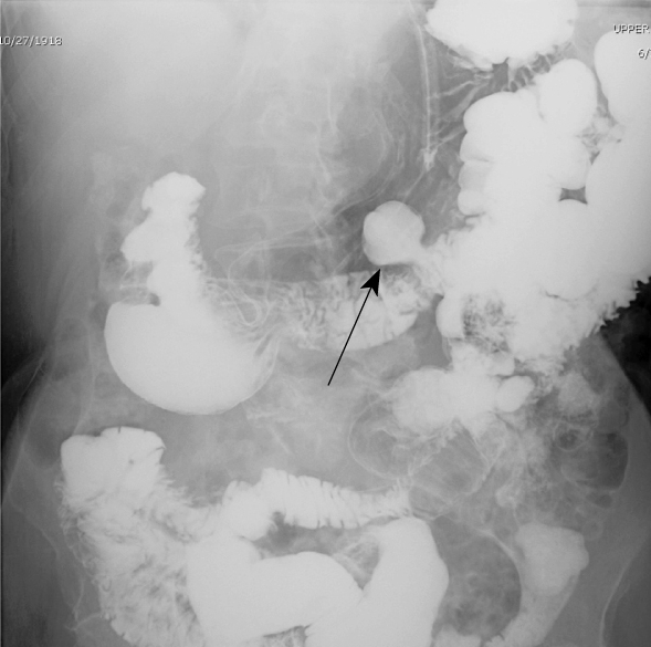
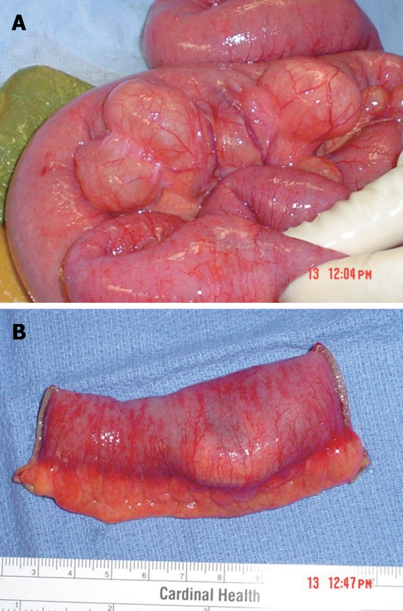
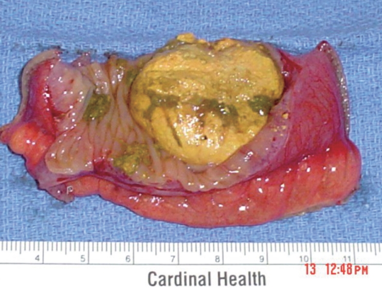
Jejunal diverticula are quite rare. Furthermore, small bowel diverticular disease resulting in enteroliths can lead to complications necessitating surgical intervention. In this manuscript, we report two presentations of jejunal diverticulum with complications from enteroliths followed by a review of the literature. The first case was that of a 79-year-old male who presented with abdominal pain and was found, on computed tomography (CT) scan, to have evidence of intestinal perforation. A laparotomy showed that he had perforated jejunal diverticulitis. The second case was that of an 89-year-old female who presented with recurrent episodes of bowel obstruction. A laparotomy showed that she had an enterolith impacted in her jejunum in the presence of significant diverticular disease. Although a rare entity, familiarity with jejunal diverticular disease, its complications, and its management, should be part of every surgeon's base of knowledge when considering abdominal pathology.
Keywords: Acute abdomen; Bowel obstruction; Diverticula disease; Enterolith; Jejunum.
Figures
References
-
- Cooper SA. The Anatomy and surgical treatment of abdominal hernia. Philadelphia: Lea and Blanchard; 1844.
-
- Silen W. Cope's Early Diagnosis of the Acute Abdomen, 21st ed. New York: Oxford University Press; 2005.
-
- Chiu EJ, Shyr YM, Su CH, Wu CW, Lui WY. Diverticular disease of the small bowel. Hepatogastroenterology. 2000;47:181–184. - PubMed
LinkOut - more resources
Full Text Sources
