A review on dural tail sign
- PMID: 21161034
- PMCID: PMC2999017
- DOI: 10.4329/wjr.v2.i5.188
A review on dural tail sign
Abstract
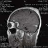
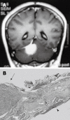
"Dural tail sign" (DTS) which is a thickening of the dura adjacent to an intracranial pathology on contrast-enhanced T1 MR Images, was first thought to be pathognomonic of meningioma, however, many subsequent studies demonstrated this sign adjacent to various intra- and extra-cranial pathologies and in spinal lesions. In this paper we outline the history, accompanying pathologies and the differentiation and probable pathophysiology of DTS. We also discuss whether we can predict tumoral involvement of the dural tail before surgery and whether the dural tail adjacent to a tumor should be resected.
Keywords: Dural tail sign; Histopathology; Magnetic resonance imaging; Meningioma.
Figures


Similar articles
-
Application of contrast-enhanced T1-weighted MRI-based 3D reconstruction of the dural tail sign in meningioma resection.J Neurosurg. 2016 Jul;125(1):46-52. doi: 10.3171/2015.5.JNS142510. Epub 2015 Dec 11. J Neurosurg. 2016. PMID: 26654184
-
Prevalence of "dural tail sign" in patients with different intracranial pathologies.Eur J Radiol. 2006 Oct;60(1):42-5. doi: 10.1016/j.ejrad.2006.04.003. Epub 2006 May 3. Eur J Radiol. 2006. PMID: 16675180
-
Pathologic significance of the "dural tail sign".Eur J Radiol. 2009 Apr;70(1):10-6. doi: 10.1016/j.ejrad.2008.01.010. Epub 2008 Feb 21. Eur J Radiol. 2009. PMID: 18294796
-
The dural tail sign--beyond meningioma.Clin Radiol. 2005 Feb;60(2):171-88. doi: 10.1016/j.crad.2004.01.019. Clin Radiol. 2005. PMID: 15664571 Review.
-
"Dural tail" adjacent to a giant posterior cerebral artery aneurysm: case report and review of the literature.Neuroradiology. 1997 Aug;39(8):577-80. doi: 10.1007/s002340050470. Neuroradiology. 1997. PMID: 9272495 Review.
Cited by
-
Dural Tail Sign in Meningiomas.AACE Clin Case Rep. 2021 Jan 13;7(3):226-227. doi: 10.1016/j.aace.2020.12.014. eCollection 2021 May-Jun. AACE Clin Case Rep. 2021. PMID: 34095494 Free PMC article. No abstract available.
-
Pneumosinus dilatans in anterior skull base meningiomas.Neuroradiology. 2013 Feb;55(3):307-11. doi: 10.1007/s00234-012-1106-9. Epub 2012 Nov 6. Neuroradiology. 2013. PMID: 23129016
-
Gamma Knife radiosurgery for intracranial meningiomas: Do we need to treat the dural tail? A single-center retrospective analysis and an overview of the literature.Surg Neurol Int. 2014 Sep 5;5(Suppl 8):S391-5. doi: 10.4103/2152-7806.140192. eCollection 2014. Surg Neurol Int. 2014. PMID: 25289168 Free PMC article.
-
Adjuvant multimodal treatment for spinal intradural extramedullary capillary hemangioma with subpial growth: illustrative case.J Neurosurg Case Lessons. 2023 Apr 10;5(15):CASE2314. doi: 10.3171/CASE2314. Print 2023 Apr 10. J Neurosurg Case Lessons. 2023. PMID: 37039294 Free PMC article.
-
Dural tail sign positive tumors: Points to make a differential diagnosis.Radiol Case Rep. 2023 Dec 4;19(2):773-779. doi: 10.1016/j.radcr.2023.10.054. eCollection 2024 Feb. Radiol Case Rep. 2023. PMID: 38089139 Free PMC article.
References
-
- Liu WC, Choi G, Lee SH, Han H, Lee JY, Jeon YH, Park HS, Park JY, Paeng SS. Radiological findings of spinal schwannomas and meningiomas: focus on discrimination of two disease entities. Eur Radiol. 2009;19:2707–2715. - PubMed
-
- Wilms G, Lammens M, Marchal G, Van Calenbergh F, Plets C, Van Fraeyenhoven L, Baert AL. Thickening of dura surrounding meningiomas: MR features. J Comput Assist Tomogr. 1989;13:763–768. - PubMed
-
- Goldsher D, Litt AW, Pinto RS, Bannon KR, Kricheff II. Dural "tail" associated with meningiomas on Gd-DTPA-enhanced MR images: characteristics, differential diagnostic value, and possible implications for treatment. Radiology. 1990;176:447–450. - PubMed
-
- Guermazi A, Lafitte F, Miaux Y, Adem C, Bonneville JF, Chiras J. The dural tail sign--beyond meningioma. Clin Radiol. 2005;60:171–188. - PubMed
-
- Rokni-Yazdi H, Sotoudeh H. Prevalence of "dural tail sign" in patients with different intracranial pathologies. Eur J Radiol. 2006;60:42–45. - PubMed
LinkOut - more resources
Full Text Sources

