. 2011 Apr;22(3-4):156-69.
doi: 10.1007/s00335-010-9309-z. Epub 2010 Dec 15.

Molecular characterization of an allelic series of mutations in the mouse Nox3 gene

Affiliations

Molecular characterization of an allelic series of mutations in the mouse Nox3 gene

John P Flaherty et al. Mamm Genome. 2011 Apr.

Abstract

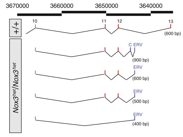
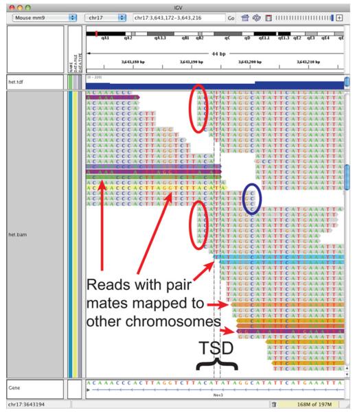
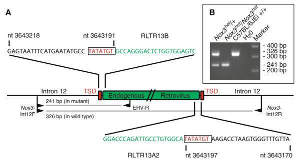
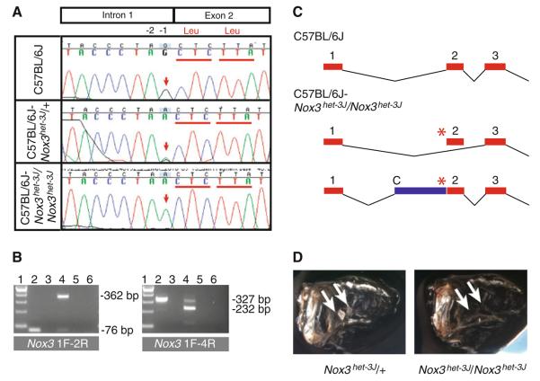
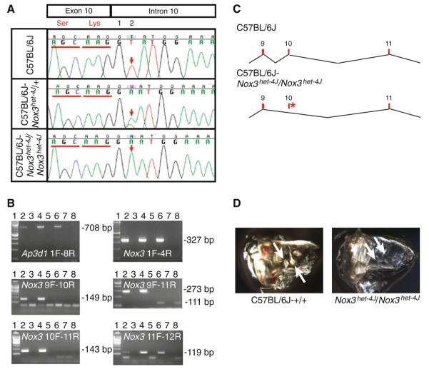
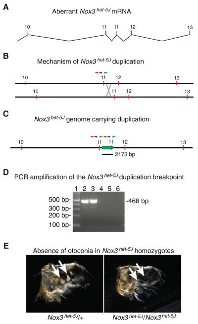
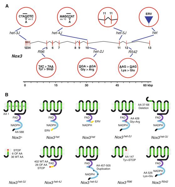
The inner ear consists of the cochlea (the organ of hearing) and the vestibular system (the organs of balance). Within the vestibular system, linear acceleration and gravity are detected by the saccule and utricle. Resting above the neurosensory epithelia of these organs are otoconia, minute proteinaceous and crystalline (calcite) inertial masses that shift under the physical forces imparted by linear movements and gravity. It is the transduction and sensation of these movements and their integration with vision and proprioceptive inputs that contribute to the sensation of balance. It has been proposed that a reactive oxygen species- (ROS-) generating NADPH oxidase comprising the gene products of the Nox3, Noxo1, and Cyba genes plays a critical and constructive role in the process of inner-ear development, specifically, the deposition of otoconia. Inactivation in mouse of any of the NADPH oxidase components encoded by the Nox3, Noxo1, or Cyba gene results in the complete congenital absence of otoconia and profound vestibular dysfunction. Here we describe our use of PCR, reverse transcription-PCR (RT-PCR), and rapid amplification of cDNA ends (RACE) with traditional and high-throughput (HTP) sequencing technologies to extend and complete the molecular characterization of an allelic series of seven mutations in the Nox3 gene. Collectively, the mutation spectrum includes an endogenous retrovirus insertion, two missense mutations, a splice donor mutation, a splice acceptor mutation, premature translational termination, and a small duplication. Together, these alleles provide tools to investigate the mechanisms of otoconial deposition over development, throughout aging, and in various disease states.


Figures

Fig. 1
Fig. 1
3′ RACE analysis of Nox3het transcripts. 3′ RACE analysis of Nox3het/Nox3het inner-ear total RNA reveals the aberrant splicing products shown here that terminate in a sequence showing similarity to the sequences of mouse endogenous retroviruses. Alternating black lines with numbers, genome coordinates on Chromosome 17 (build 37); top schematic, 3′ RACE products from wild-type RNA; remaining schematics, 3′ RACE products from mutant RNA that splice into an exon derived from the endogenous retroviral sequence (ERV), may include a cryptic exon (C) or may skip exons 11 and 12. The size difference observed between the second and third mutants' 3′ RACE products is due to a difference in the size of included ERV sequence
Fig. 2
Fig. 2
Integrative genome viewer (IGV) display of the endogenous retroviral insertion site in the Nox3het allele. The IGV display of the endogenous retroviral insertion site reveals signatures indicative of the insertion, including truncation of reads directed distally (red ovals), truncation of reads directed proximally (blue oval), reads with pair mates mapped to other chromosomes (variously colored reads), and overlap of truncated reads in the area of an ACATATA target site duplication (TSD)
Fig. 3
Fig. 3
Detailed description of the endogenous retroviral insertion site in the Nox3het allele. a Schematic describing the endogenous retroviral (ERV) insertion in the Nox3het allele. Central horizontal line, intron 12 of the Nox3 gene; green rectangle, ERV; red rectangles, target site duplications (TSD); black sequences, partial sequence from intron 12; red sequences, target site duplication; green sequences, partial ERV sequences showing similarity to ERV classes RLTR13B and RLTR13A2; nucleotide (nt) numbers, Chromosome 17 genome positions (build 37); black flags, PCR primers; gray lines, amplification products from the wild-type and mutant alleles. b (inset) The Nox3het genotyping assay detects a 326-bp fragment in wild-type mice, a 241-bp fragment in Nox3het mutant mice, and both fragments in heterozygous mice
Fig. 4
Fig. 4
Analysis of the Nox3het–3J allele. a Sanger sequencing-based confirmation of the Nox3het–3J mutation identified by HTP sequencing. The mutation consists of a G > A transition at the −1 position of the splice acceptor site just prior to exon 2. b RT-PCR analysis of C57BL/6J and Nox3het–3J/Nox3het–3J inner-ear RNA with 1F/2R and 1F/4R primer pairs. Lane 1, 100-bp ladder; lane 2, wild-type RNA; lane 3, negative reverse transcriptase (−RT) control; lane 4, Nox3het–3J/Nox3het–3J RNA; lane 5, −RT control; lane 6, no template control. c Models of wild-type (C57BL/6J) and mutant (C57BL/6J-Nox3het–3J/Nox3het–3J) Nox3 transcripts. Red numbered rectangles, exons; asterisk, mutation; C, cryptic splice acceptor; blue rectangle, intronic sequence incorporated into the mutant transcript. d Whole-mount preparation of cleared temporal bones from adult Nox3het–3J/+ and Nox3het–3J/Nox3het–3J mice. Note the presence of otoconia (arrows) in the heterozygotes and their absence in mutant homozygotes
Fig. 5
Fig. 5
Analysis of the Nox3het–4J allele. a Sanger sequencing-based identification of the Nox3het–4J mutation. The mutation consists of a T > A transversion at the +2 position of the splice donor site just after exon 10. b RT-PCR analysis of C57BL/6J and Nox3het–4J/Nox3het–4J inner-ear RNA with Ap3d1 1F/8R (positive control) primers as well as the Nox3 1F/4R, 9F/10R, 9F/11R, 10F/11R, and 11F/12R primer pairs. Lane 1, 100-bp ladder; lane 2, wild-type RNA; lane 3, negative reverse transcriptase (−RT) control; lane 4, Nox3het–4J/+ RNA; lane 5, −RT control; lane 6, Nox3het–4J/Nox3het–4J RNA; lane 7, −RT control; lane 8, no template control. c Models of wild-type (C57BL/6J) and mutant (C57BL/6J-Nox3het–4J/Nox3het–4J) Nox3 transcripts. Red numbered rectangles, exons; asterisk, mutation. d Whole-mount preparation of cleared temporal bones from adult C57BL/6J-+/+and Nox3het–4J/Nox3het–4J mice. Note the presence of otoconia (arrows) in the wild-type preparation and their absence in mutant homozygotes
Fig. 6
Fig. 6
Analysis of the Nox3het–5J allele. a RT-PCR analysis and Sanger sequencing identified a duplication of exon 11 in Nox3het–5J cDNA. Numbered red rectangles, exons. b Proposed mechanism for the Nox3het–5J duplication by uneven crossing over. Numbered red rectangles, exons; red triangle, Nox3-44516R primer; blue triangle, het-5JR primer; green triangle, Nox3-46393F primer; gray X, crossover event. c Schematic of the Nox3het–5J allele. Red numbered rectangles, exons; green rectangle, duplicated region of 2173 bp encompassing exon 11; red, blue, and green triangles are as in b. Note that in the duplication-bearing allele, the three primers located between exons 11 are now pointed toward each and are able to support amplification. d Amplification across the duplication in the Nox3het–5J allele with the Nox3-46393F and het-5JR primers. Lane 1, 100-bp ladder; lanes 2–3, Nox3het–5J/Nox3het–5J DNA; lanes 4–5, wild-type DNA; lane 6, no template control. e Whole-mount preparation of cleared temporal bones from adult Nox3het–5J/+ and Nox3het–5J/Nox3het–5J mice. Note the presence of otoconia (arrows) in the heterozygotes and their absence in mutant homozygotes
Fig. 7
Fig. 7
A summary of the mutations in Nox3 and their effects on the NOX3 protein. a Shown are the Nox3 transcription unit (numbered vertical red lines, exons) as well as the mutations occurring in the het, het-2J, het-3J, het-4J, het-5J, R96, and R542 alleles of Nox3. For the het allele: ERV endogenous retrovirus. For the het-2J, R96, and R542 alleles: shown are the wild-type (left) and mutant (right) codons and amino acids. The nucleotide that underwent mutation is underlined. For the het-3J and het-4J alleles: shown are seven nucleotides at the intron/exon boundary. Gray labeled rectangle, exon; underlined, wild-type nucleotide with mutant nucleotide shown below; overlined, AG/GT splicing acceptor/donor dinucleotide sequence. For the het-5J allele: shown are the duplicated exons 11. Also shown is a 60-kbp scale. b Shown schematically are the predicted effects of Nox3 mutant alleles on the NOX3 protein. Numbered green vertical lines, transmembrane domains; purple lines, FAD binding domains; blue lines, NADPH binding domains; yellow lines, novel amino acids encoded by the endogenous retrovirus (het) or due to frameshift mutations (het-3J and het-4J); red asterisks, missense mutations; shortened first transmembrane domain in het-3J, in-frame deletion; red lines, premature stop codons; segmented blue line in het-5J, in-frame duplication of the second region of the NADPH binding domain; AA amino acid, ERV endogenous retrovirus, FAD, FAD binding domain; NADPH, NADPH binding domain; OF out-of-frame, WT wild type. This figure is a schematic representation of the effects of Nox3 mutant alleles on translation and is not meant to imply that mutant proteins are present or properly targeted to the cell membrane

References

    1. Banfi B, Malgrange B, Knisz J, Steger K, Dubois-Dauphin M, et al. NOX3, a superoxide-generating NADPH oxidase of the inner ear. J Biol Chem. 2004;279:46065–46072. - PubMed
    1. Bedard K, Krause KH. The NOX family of ROS-generating NADPH oxidases: physiology and pathophysiology. Physiol Rev. 2007;87:245–313. - PubMed
    1. Bergstrom DE, Bergstrom RA, Munroe RJ, Schimenti JC, Gagnon LH, et al. The Nox3 and Noxo1 genes, encoding presumptive members of an NADPH oxidase complex, are required for normal vestibular function and development; 18th international mammalian genome conference; Seattle, WA. 17–22 Oct 2004.2004.
    1. Burke B, Stewart CL. The laminopathies: the functional architecture of the nucleus and its contribution to disease. Annu Rev Genomics Hum Genet. 2006;7:369–405. - PubMed
    1. Capell BC, Collins FS. Human laminopathies: nuclei gone genetically awry. Nat Rev Genet. 2006;7:940–952. - PubMed

Publication types