Review
. 2011 Mar;68(5):817-31.
doi: 10.1007/s00018-010-0571-8. Epub 2010 Dec 14.

Emerging knowledge of regulatory roles of D-amino acids in bacteria

Affiliations
Review

Emerging knowledge of regulatory roles of D-amino acids in bacteria

Felipe Cava et al. Cell Mol Life Sci. 2011 Mar.

Abstract

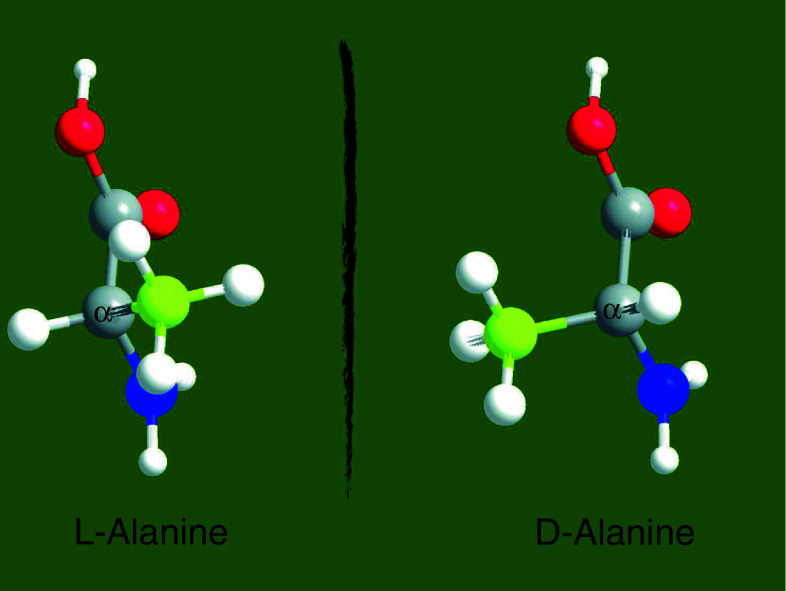
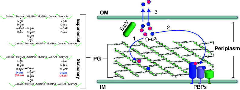
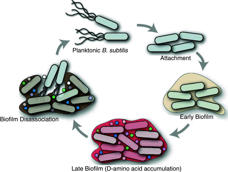
The D-enantiomers of amino acids have been thought to have relatively minor functions in biological processes. While L-amino acids clearly predominate in nature, D-amino acids are sometimes found in proteins that are not synthesized by ribosomes, and D-Ala and D-Glu are routinely found in the peptidoglycan cell wall of bacteria. Here, we review recent findings showing that D-amino acids have previously unappreciated regulatory roles in the bacterial kingdom. Many diverse bacterial phyla synthesize and release D-amino acids, including D-Met and D-Leu, which were not previously known to be made. These noncanonical D-amino acids regulate cell wall remodeling in stationary phase and cause biofilm dispersal in aging bacterial communities. Elucidating the mechanisms by which D-amino acids govern cell wall remodeling and biofilm disassembly will undoubtedly reveal new paradigms for understanding how extracytoplasmic processes are regulated as well as lead to development of novel therapeutics.


Figures

Fig. 1
Fig. 1
Chirality of alanine. Ball-and-sticks representation of the enantiomeric forms of the amino acid alanine. Carboxyl group is colored in red, amino group in blue, and R-group in yellow. Chiral carbon is labeled as α. The molecules were designed with ChemDraw Ultra 12.0 and Chem3D Pro 12.0 software
Fig. 2
Fig. 2
Biosynthesis of peptidoglycan (PG). General scheme of PG synthesis in gram negative bacteria. PG synthesis is initiated with the synthesis of the disaccharide pentapeptide precursors in the cytosol (GlcNAc-MurNAc-l-Ala-d-Glu-DAP-d-Ala-d-Ala) [42]. Then, PG precursors are translocated to the periplasmic space facilitated by the formation of lipidic complexes with bactoprenol [43, 44]. Once in the periplasm, PG monomers are incorporated into the murein polymer by transglycosylation and transpeptidation reactions carried out by the activity of the penicillin-binding proteins (PBPs) [–48]. Also PBP activities (murein hydrolases) can affect the length of the stem peptides, depicted as d-Ala between brackets [43]. IM Inner membrane, OM outer membrane
Fig. 3
Fig. 3
Mechanism of amino acid racemase reactions. a PLP-dependent alanine racemase (AlaRac) and b PLP-independent glutamate racemase (GluRac) reactions are depicted showing the amino acidic residues of the enzymes that participate in the stereochemical transformation of alanine and glutamic acid, respectively. The achiral anionic quinoid intermediate is shown in brackets. c Schematic of the catalytic mechanism of the PLP-dependent alanine racemase reaction. The molecules were designed with ChemDraw Ultra 12.0 software
Fig. 4
Fig. 4
Model of PG remodeling governed by d-amino acid release in stationary phase. PG in V. cholerae is composed of linear glycan strands made up of repeating disaccharide units of GlcNAc and MurNAc cross-linked by short peptides that consist of l-Ala, d-Glu, meso-diaminopimelic acid (m-DAP), and d-Ala. In stationary phase, d-Met (blue circles) and d-Leu (red circles) are produced by BsrV, a periplasmic racemase. These d-amino acids (1) are incorporated at the 4th position of the PG-peptide bridge where d-Ala is usually found, (2) regulate the activity of periplasmic enzymes including penicillin-binding proteins (PBPs), which synthesize and modify PG, and (3) are released into the extracellular milieu where d-amino acids regulate the PG of other bacteria. OM Outer membrane, IM inner membrane
Fig. 5
Fig. 5
Model of biofilm life cycle. B. subtilis cells associated in biofilm communities produce d-amino acids (d-Tyr, d-Met, d-Trp, and d-Leu represented with colored circles), which accumulate and trigger biofilm dispersal

References

    1. Bentley R. Chiral: a confusing etymology. Chirality. 2010;22:1–2. doi: 10.1002/chir.20699. - DOI - PubMed
    1. Flack HD. Louis Pasteur’s discovery of molecular chirality and spontaneous resolution in 1848, together with a complete review of his crystallographic and chemical work. Acta Crystallogr A. 2009;65:371–389. doi: 10.1107/S0108767309024088. - DOI - PubMed
    1. Gal J. The discovery of biological enantioselectivity: Louis Pasteur and the fermentation of tartaric acid, 1857—a review and analysis 150 yr later. Chirality. 2008;20:5–19. doi: 10.1002/chir.20494. - DOI - PubMed
    1. Meierhenrich U. Stereochemistry for the study of the origin of life. Heidelberg: Springer; 2008.
    1. Kondepudi DK, Kaufman RJ, Singh N. Chiral symmetry breaking in sodium chlorate crystallization. Science. 1990;250:975–976. doi: 10.1126/science.250.4983.975. - DOI - PubMed

Publication types

LinkOut - more resources