Comparative Study
. 2011 Mar;16(3):221-34.
doi: 10.1007/s10495-010-0566-9.

Cell death-resistance of differentiated myotubes is associated with enhanced anti-apoptotic mechanisms compared to myoblasts

Affiliations
Comparative Study

Cell death-resistance of differentiated myotubes is associated with enhanced anti-apoptotic mechanisms compared to myoblasts

Rijin Xiao et al. Apoptosis. 2011 Mar.

Abstract

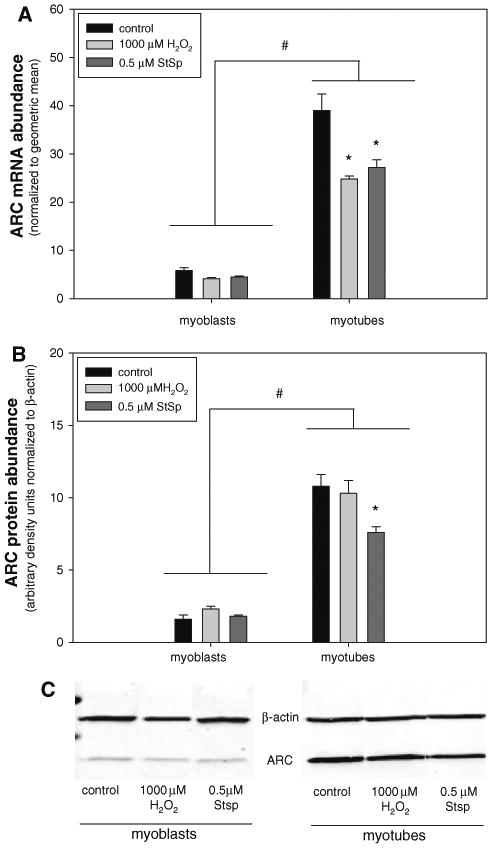
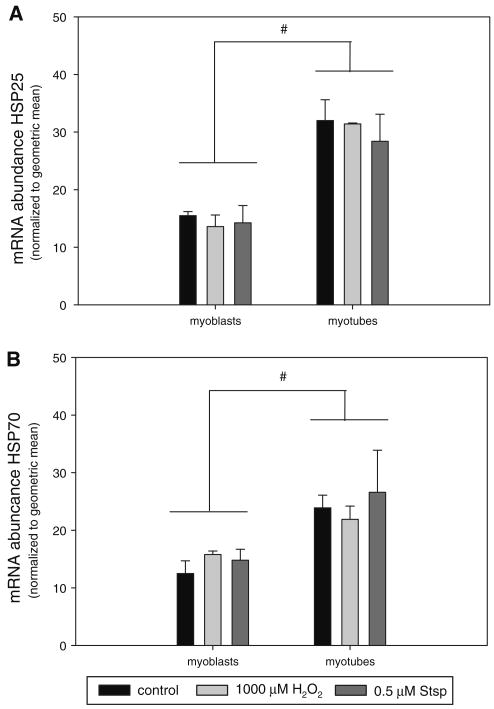
Skeletal muscle atrophy is associated with elevated apoptosis while muscle differentiation results in apoptosis resistance, indicating that the role of apoptosis in skeletal muscle is multifaceted. The objective of this study was to investigate mechanisms underlying apoptosis susceptibility in proliferating myoblasts compared to differentiated myotubes and we hypothesized that cell death-resistance in differentiated myotubes is mediated by enhanced anti-apoptotic pathways. C(2)C(12) myoblasts and myotubes were treated with H(2)O(2) or staurosporine (Stsp) to induce cell death. H(2)O(2) and Stsp induced DNA fragmentation in more than 50% of myoblasts, but in myotubes less than 10% of nuclei showed apoptotic changes. Mitochondrial membrane potential dissipation was detected with H(2)O(2) and Stsp in myoblasts, while this response was greatly diminished in myotubes. Caspase-3 activity was 10-fold higher in myotubes compared to myoblasts, and Stsp caused a significant caspase-3 induction in both. However, exposure to H(2)O(2) did not lead to caspase-3 activation in myoblasts, and only to a modest induction in myotubes. A similar response was observed for caspase-2, -8 and -9. Abundance of caspase-inhibitors (apoptosis repressor with caspase recruitment domain (ARC), and heat shock protein (HSP) 70 and -25 was significantly higher in myotubes compared to myoblasts, and in addition ARC was suppressed in response to Stsp in myotubes. Moreover, increased expression of HSPs in myoblasts attenuated cell death in response to H(2)O(2) and Stsp. Protein abundance of the pro-apoptotic protein endonuclease G (EndoG) and apoptosis-inducing factor (AIF) was higher in myotubes compared to myoblasts. These results show that resistance to apoptosis in myotubes is increased despite high levels of pro-apoptotic signaling mechanisms, and we suggest that this protective effect is mediated by enhanced anti-caspase mechanisms.


Conflict of interest statement

Conflict of interest: The authors declare that they have no conflict of interest.

Figures

Fig. 1
Fig. 1
C2C12 differentiation and apoptosis. Representative picture of fully differentiated C2C12 myotubes after 72 h in differentiation medium (a). Myogenin gene expression of myoblast and differentiated myotubes (b). Representative pictures of untreated control C2C12 myoblasts (c), myoblasts undergoing apoptosis induced by 1000 μM H2O2 (d) or 0.5 μM Stsp (e). Values are means ± SE. * Indicates a significant difference from control (P < 0.05)
Fig. 2
Fig. 2
Myotubes are more resistant to apoptosis than myoblasts. Myoblasts and myotubes were exposed to H2O2 (ad) or Stsp (e, f) for 3 h, and the frequency of TUNEL-positive nuclei was counted and displayed (b, d, e, f). Representative pictures of myoblasts (a) and myotubes (c) after treatment with 1000 μM H2O2 indicating a lower number of TUNEL positive nuclei in myotubes. Percentage of TUNEL positive nuclei in myoblasts (b + e) and myotubes (d + f) after treatment with H2O2 (b + d) or Stsp (e + f). Results represented mean ± SE. a, b, c Indicate significant difference between treatments without same letters (P < 0.05). Note the difference in values on y-axis
Fig. 3
Fig. 3
Mitochondrial membrane potential (Δψm) transition is attenuated in myotubes. Untreated (control) or treated (1000 μM H2O2 or 0.5 μM Stsp) C2C12 myoblasts and myotubes were stained with JC-1 and observed under TRITC (590 nm) and GFP (530 nm) filters. Representative pictures are shown in top of figure. Δψm index (represented as the ratio of red to green fluorescence) of control (black bars), 1000 μM H2O2 (light grey bars) or 0.5 μM Stsp (dark grey bars) in myoblasts and myotubes. Values are mean ± SE. # Indicates a significant main effect between myoblasts and myotubes. * Indicates a significant difference compared to control within the cell type (P < 0.05)
Fig. 4
Fig. 4
High level of caspase activities in differentiated myotubes. Caspase -2, -3, -8 and -9 activities of myoblasts (black bars) and myotubes (grey bars) are depicted. Values are mean ± SE. * Indicates a significant difference compared to myoblasts (P < 0.05)
Fig. 5
Fig. 5
Caspase activation in myoblasts and myotubes following Stsp treatment. Caspase-3 (a), -2 (b), -8 (c) and -9 (d) activities in myoblasts and myotubes treated with H2O2 and Stsp. Units of H2O2 and Stsp are in μM. Values are mean ± SE. * Indicates a significant difference of Stsp compared to corresponding controls. ˆ Indicates a significant difference of H2O2 compared to corresponding controls (P < 0.05)
Fig. 6
Fig. 6
ARC gene expression is highly elevated in myotubes. mRNA (a) and protein abundance (b) and representative immunoblot of Western analysis (c) of ARC in myoblasts and myotubes in response to 1000 μM H2O2 (light grey bars) or 0.5 μM Stsp (dark grey bars) is depicted. Values are means ± SE. # Indicates a significant main effect for myoblasts and myotubes. * Indicates a significant difference from control within myotubes (P < 0.05)
Fig. 7
Fig. 7
Heat shock protein gene expression is elevated in differentiated myotubes. mRNA abundance of HSP27 (a) and HSP70 (b) in myoblasts and myotubes under control conditions (black bars) and after treatment with 1000 μM H2O2 (light grey bars) or 0.5 μM Stsp (dark grey bars) is depicted. Values are means ± SE. # Indicates a significant main effect between myoblasts and myotubes (P < 0.05)
Fig. 8
Fig. 8
Elevated HSP expression enhances cell survival. Representative immunoblots of HSP27 and HSP70 in myoblasts at 37°C and 42°C and of myotubes at 37°C are depicted and compared to β-actin (a). Cell survival of myoblasts at 37°C (black bars) or 42°C (grey bars) under control conditions or after treatment with 1000 μM H2O2 (H2O2) or 0.5 μM Stsp (Stsp) is shown (b). Values are means ± SE. * Indicates significant main effect between 37°C and 42°C (P < 0.05)
Fig. 9
Fig. 9
AIF protein abundance is elevated in myotubes. AIF protein abundance normalized to β-actin in cytosolic (a) and nuclear (b) fractions of myoblasts and myotubes under control conditions (black bars) or after treatment with 0.5 μM Stsp (light grey bars) or 1000 μM H2O2 (dark grey bars). Representative AIF immunoblot (c) of cytosolic and nuclear fractions of myoblasts and myotubes under control conditions (c) or treated with 0.5 μM Stsp (S) or 1000 μM H2O2 (H). Values are means ± SE. # Indicates a significant main effect between myoblasts and myotubes (P < 0.05)
Fig. 10
Fig. 10
EndoG protein abundance is elevated in myotubes. EndoG protein abundance normalized to β-actin in cytosolic (a) and nuclear (b) fractions of myoblasts and myotubes under control conditions (black bars) or after treatment with 0.5 μM Stsp (light grey bars) or 1000 μM H2O2 (dark grey bars). Representative AIF immunoblot (c) of cytosolic and nuclear fractions of myoblasts and myotubes under control conditions (c) or treated with 0.5 μM Stsp (S) or 1000 μM H2O2 (H). Values are means ± SE. # Indicates a significant main effect between myoblasts and myotubes; * Indicates significant difference from control within cell type (P < 0.05)

Similar articles

Cited by

References

    1. Jagani Z, Khosravi-Far R. Cancer stem cells and impaired apoptosis. Adv Exp Med Biol. 2008;615:331–344. - PubMed
    1. Melet A, Song K, Bucur O, Jagani Z, Grassian AR, Khosravi-Far R. Apoptotic pathways in tumor progression and therapy. Adv Exp Med Biol. 2008;615:47–79. - PubMed
    1. Friedlander RM. Apoptosis and caspases in neurodegenerative diseases. N Engl J Med. 2003;348:1365–1375. - PubMed
    1. Kermer P, Liman J, Weishaupt JH, Bahr M. Neuronal apoptosis in neurodegenerative diseases: from basic research to clinical application. Neurodegener Dis. 2004;1:9–19. - PubMed
    1. Adams V, Jiang H, Yu J, et al. Apoptosis in skeletal myocytes of patients with chronic heart failure is associated with exercise intolerance. J Am Coll Cardiol. 1999;33:959–965. - PubMed

Publication types

MeSH terms