. 2010 Dec 16:8:134.
doi: 10.1186/1479-5876-8-134.

Development of targeted therapy for bladder cancer mediated by a double promoter plasmid expressing diphtheria toxin under the control of H19 and IGF2-P4 regulatory sequences

Affiliations

Development of targeted therapy for bladder cancer mediated by a double promoter plasmid expressing diphtheria toxin under the control of H19 and IGF2-P4 regulatory sequences

Doron Amit et al. J Transl Med. .

Abstract

Background: The human IGF2-P4 and H19 promoters are highly active in a variety of human cancers (including bladder cancer), while existing at a nearly undetectable level in the surrounding normal tissue.Single promoter vectors expressing diphtheria toxin A-fragment (DTA) under the control regulation of IGF2-P4 or H19 regulatory sequences (IGF2-P4-DTA and H19-DTA) were previously successfully used in cell lines, animal models and recently in human patients with superficial cell carcinoma of the bladder (treated with H19-DTA). However this targeted medicine approach could be limited, as not all cancer patients express high levels of H19. Hence, a double promoter DTA-expressing vector was created, carrying on a single construct two separate genes expressing the diphtheria toxin A-fragment (DTA), from two different regulatory sequences, selected from the cancer-specific promoters H19 and IGF2-P4.

Methods: H19 and IGF2-P4 gene expression was tested in samples of Transitional Cell Carcinoma (TCC) of the bladder by in-situ hybridization (ISH) and by quantitative Real-Time PCR (qRT-PCR). The therapeutic potential of the double promoter toxin vector H19-DTA-IGF2-P4-DTA was tested in TCC cell lines and in heterotopic and orthotopic animal models of bladder cancer.

Results: Nearly 100% of TCC patients highly expressed IGF2-P4 and H19, as determined by ISH and by qRT-PCR. The double promoter vector exhibited superior tumor growth inhibition activity compared to the single promoter expression vectors, in cell lines and in heterotopic and orthotopic bladder tumors.

Conclusions: Our findings show that bladder tumors may be successfully treated by intravesical instillation of the double promoter vector H19-DTA-P4-DTA.Overall, the double promoter vector exhibited enhanced anti-cancer activity relative to single promoter expression vectors carrying either gene alone.


Figures

Figure 1
Figure 1
A schematic illustration depicting the construction of the double promoter H19-DTA-P4-DTA expression vector: The coding sequence of each DTA is under the transcriptional control of both H19 and IGF2-P4 promoter sequences, respectively, Kana (R) - kanamycine resistance gene.
Figure 2
Figure 2
ISH detection of the expression of IGF2-P4 and H19 transcripts in human TCC tissue samples: IGF2-P4 (A) and H19 (B) specific transcripts, detected by ISH. The positive stained cells are marked by black arrows (Magnification are ×20).
Figure 3
Figure 3
In vitro enhanced protein synthesis inhibition activity of H19-DTA-P4-DTA in human bladder carcinoma cell lines: Tumor growth inhibition activity of the H19-DTA, P4-DTA and H19-DTA-P4-DTA vectors in T24P (A-B) and HT-1376 (C-D) cells was measured as a reduction of LucSV40 activity. Cells were cotransfected with 2 μg of LucSV40 and the indicated concentrations of the DTA expressing vectors, or with LucSV40 alone. Transfection experiments were stopped after 48 hours and luciferase activity was assessed. The decrease in LucSV40 activity was determined by comparison to the same cell type transfected with LucSV40 alone as a measure for cytotoxicity. The diverse effect of each vector at the lowest plasmid transfected concentration is indicated (B, D).
Figure 4
Figure 4
The expression of H19 and IGF2-P4 in heterotopic subcutaneous tumors determined by RT-PCR: The expression of H19 and IGF2-P4 transcripts in heterotopic subcutaneous tumors after injection of T24P (A) or HT-1376 cells (B) was determined by RT-PCR. "M": 100-bp molecular weight marker, lanes 1-2: heterotopic subcutaneous tumors from different mice induced by injection of T24P (A) or HT-1376(B) cells, lane 3: subcutaneous tissue of normal mouse, lanes 4: T24P (A) or HT-1376(B) cell lines, "C": negative control for PCR. The sizes of the PCR products are 300 bp for human H19, 119 bp for IGF2-P4 and 213 bp for Histone 3.3 internal control, respectively.
Figure 5
Figure 5
In vivo inhibition of heterotopic tumors in response to H19-DTA-P4-DTA treatments. Inhibition of tumor growth in response to H19-DTA, P4-DTA, or H19-DTA-P4-DTA treatments is shown. The tumor sizes of tumors treated with the DTA expressing vector, or with control luciferase expressing vectors were determined prior to each treatment and before sacrifice. The fold increase in tumor volume was calculated relative to the initial volume at the day of the first treatment.
Figure 6
Figure 6
Heterotopic tumors treated by H19-DTA-P4-DTA. Heterotopic bladder tumors treated with H19-DTA-P4-DTA vector (black) or with H19-Luc-P4-Luc control vector (white) were excised and the ex-vivo tumors volume were measured (A) and weighted (B). C-D: Necrosis of heterotopic tumors treated with H19-DTA-P4-DTA: Hematoxylin Eosin (HE) staining (×10) of representative sections of tumors treated with H19-Luc-P4-Luc (C), or with H19-DTA-P4-DTA (D). The necrotic areas are indicated by arrows (D). Inserts are macroscopic photographs of the heterotopic tumors.
Figure 7
Figure 7
Orthotopic bladder tumors kinetics, 14 days after intravesical cells instillation: A). "M": 100-bp molecular weight marker, lanes 1-3: orthotopic bladder tumors from different mice induced by intravesical instillation of 10 × 106 T24P cells, lane 4: bladder of normal mouse, "c": negative control for PCR. B). HE staining (×10) of a representative section of orthotopic bladder (14 days after intravesical inoculation of 10 × 106 T24P cells). The tumor area is indicated (by green line). ('U', urothelium, 'LP', lamina propria, 'M', muscularis).
Figure 8
Figure 8
The effect of intravesical treatment with H19-DTA-P4-DTA vector in orthotopic bladder carcinoma: Orthotopic tumors were induced by intravesical instillation of T24P cells, in nude mice bladders. 7 days later, mice of each group (n = 6) received an intravesical treatment with 20 μg of H19-DTA-P4-DTA, or H19-Luc-P4-Luc for each mouse. The same treatments were repeated after 3 days, and 4 days later mice were sacrificed. The bladders of both groups were excised, weighted, and the area of the malignant tissue of each bladder was determined by ImagePro Plus software. Another 4 healthy mice were used as control. The total tumor area of each bladder was determined and the mean of the total areas was calculated for each group. The Mean and SD of bladder tumor area (A) and weight (B) are shown.
Figure 9
Figure 9
Macroscopic and histopathological views of the orthotopic bladders treated with H19-DTA-P4-DTA: Shown are macroscopic photographs of the whole orthotopic bladders treated with H19-Luc-P4-Luc (A), or with H19-DTA-P4-DTA (D). The bladders of both of the groups were excised, and the area of the malignant tissue of each bladder is indicated (by grin line) for the H19-Luc-P4-Luc (B) and H19-DTA-P4-DTA (E). Histopathological microscopic view (H&E × 10 is shown for H19-Luc-P4-Luc treated bladder (C), or with H19-DTA-P4-DTA treated bladder (F) and the tumor areas are indicated (by green line), ('U', urothelium,'LP', lamina propria, 'M', muscle).
Figure 10
Figure 10
Detection of DTA and Luc transcripts in orthotopic bladder tumors: Mice with heterotopic bladder tumors were intravesically treated twice in 3 days interval, and were sacrificed 4 days after the last treatment. Tumors were excised and frozen immediately and 400 ng RNA (extracted from the tumors) was used for determination of luciferase and DTA by RT-PCR reaction. A). tumors treated with H19-Luc-P4-Luc (lanes 1-2), or with H19-DTA-P4-DTA (lanes 3-4). Lane 5: untreated orthotopic bladder tumor, 'C': negative control for PCR, 'M': 100 bp DNA ladder. The sizes of the PCR products are 468 bp and 328 bp, for DTA and Luc respectively. The lower panel shows the histone 3.3 internal control. Necrosis of orthotopic bladder tumor treated with H19-DTA-P4-DTA (H&E × 20) is shown (B) and the necrotic area is indicated (by green line). ('U', urothelium, 'LP', lamina propria, 'M', muscle).
Figure 11
Figure 11
Enhanced activity of H19-DTA-P4-DTA in human bladder carcinoma cell lines: The protein synthesis inhibition activity of the H19-DTA-P4-DTA vector in T24P (A-B) and HT-1376 (C-D) cells was measured as a reduction of LucSV40 activity, and was compared to the combination activity of H19-DTA + P4-DTA. Cells were cotransfected with 2 μg of LucSV40, and with the indicated concentrations of the DTA expressing vectors or LucSV40 alone. Transfection experiments were stopped after 48 hours and luciferase activity was assessed. The decrease in LucSV40 activity was determined by comparison to the same cell type transfected with LucSV40 alone as a measure for cytotoxicity. Enhanced effect of H19-DTA-P4-DTA vector at the lowest plasmid transfected concentration (0.005 μg compared to 0.005 μg + 0.005 μg of the combination transfection of both vectors H19-DTA + P4-DTA) is indicated (B, D).
Figure 12
Figure 12
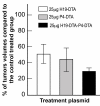
Augmented-than-additive activity of H19-DTA-P4-DTA in heterotopic bladder tumors, induced by T24P cells. The inhibition of heterotopic bladder tumor growth, induced by T24P cells is indicated by the fold increase of each DTA mice treated group compared to the control Luc treated mice. Shown are tumors treated with: 25 μg of H19-DTA, 25 μg of P4-DTA, 25 μg of H19-DTA + 25 μg of P4-DTA 25 μg of H19-DTA-P4-DTA and 50 μg of H19-DTA-P4-DTA.

References

    1. Marston LW, Zbar B, Leach F, Cordon-Cardo C, Issacs W. In: Cancer: Principles & practice of oncology. VTDeVita, SHellman, SARosenberg, editor. Philadelppia: Lippincott Williams & Wilkins; 2001. Cancer of the genitourinary system; pp. 1343–1489.
    1. Matouk IJ, DeGroot N, Mezan S, Ayesh S, Abu-lail R, Hochberg A, Galun E. The H19 non-coding RNA is essential for human tumor growth. PLoS ONE. 2007;2:e845. doi: 10.1371/journal.pone.0000845. - DOI - PMC - PubMed
    1. Matouk IJ, Mezan S, Mizrahi A, Ohana P, Abu-Lail R, Fellig Y, Degroot N, Galun E, Hochberg A. The oncofetal H19 RNA connection: hypoxia, p53 and cancer. Biochim Biophys Acta. 2010;1803:443–451. doi: 10.1016/j.bbamcr.2010.01.010. - DOI - PubMed
    1. Yao X, Hu JF, Daniels M, Shiran H, Zhou X, Yan H, Lu H, Zeng Z, Wang Q, Li T, Hoffman AR. A methylated oligonucleotide inhibits IGF2 expression and enhances survival in a model of hepatocellular carcinoma. J Clin Invest. 2003;111:265–273. - PMC - PubMed
    1. Ariel I, Lustig O, Schneider T, Pizov G, Sappir M, De-Groot N, Hochberg A. The imprinted H19 gene as a tumor marker in bladder carcinoma. Urology. 1995;45:335–338. doi: 10.1016/0090-4295(95)80030-1. - DOI - PubMed

MeSH terms