

Foot posture in people with medial compartment knee osteoarthritis
- PMID: 21162748
- PMCID: PMC3020154
- DOI: 10.1186/1757-1146-3-29
Foot posture in people with medial compartment knee osteoarthritis
Abstract
Background: Foot posture has long been considered to contribute to the development of lower limb musculoskeletal conditions as it may alter the mechanical alignment and dynamic function of the lower limb. This study compared foot posture in people with and without medial compartment knee osteoarthritis (OA) using a range of clinical foot measures. The reliability of the foot measures was also assessed.
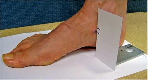
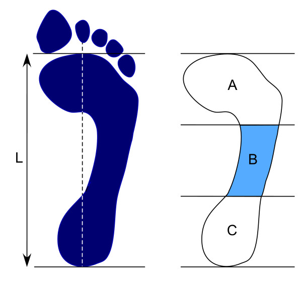
Methods: The foot posture of 32 patients with clinically and radiographically-confirmed OA predominantly in the medial compartment of the knee and 28 asymptomatic age-matched healthy controls was investigated using the foot posture index (FPI), vertical navicular height and drop, and the arch index. Independent t tests and effect size (Cohen's d) were used to investigate the differences between the groups in the foot posture measurements.
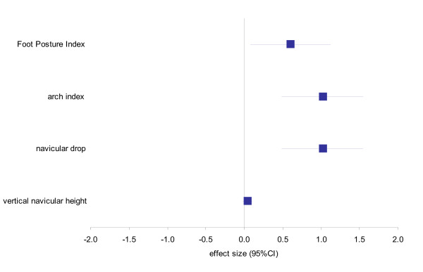
Results: Significant differences were found between the control and the knee OA groups in relation to the FPI (1.35 ± 1.43 vs. 2.46 ± 2.18, p = 0.02; d = 0.61, medium effect size), navicular drop (0.02 ± 0.01 vs. 0.03 ± 0.01, p = 0.01; d = 1.02, large effect size) and the arch index (0.22 ± 0.04 vs. 0.26 ± 0.04, p = 0.04; d = 1.02, large effect size). No significant difference was found for vertical navicular height (0.24 ± 0.03 vs. 0.23 ± 0.03, p = 0.54; d = 0.04, negligible effect size).
Conclusion: People with medial compartment knee OA exhibit a more pronated foot type compared to controls. It is therefore recommended that the assessment of patients with knee OA in clinical practice should include simple foot measures, and that the potential influence of foot structure and function on the efficacy of foot orthoses in the management of medial compartment knee OA be further investigated.
Figures
References
-
- Access Economics. Painful realities: the economic impact of arthritis in Australia in 2007. Sydney: Arthritis Australia; 2007.
LinkOut - more resources
Full Text Sources
