. 2011 Jan;49(1):56-65.
doi: 10.1016/j.ceca.2010.11.008. Epub 2010 Dec 15.

Inhibition of spontaneous activity of rabbit atrioventricular node cells by KB-R7943 and inhibitors of sarcoplasmic reticulum Ca(2+) ATPase

Affiliations

Inhibition of spontaneous activity of rabbit atrioventricular node cells by KB-R7943 and inhibitors of sarcoplasmic reticulum Ca(2+) ATPase

Hongwei Cheng et al. Cell Calcium. 2011 Jan.

Abstract

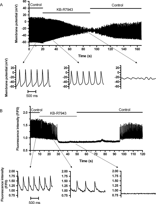
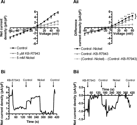
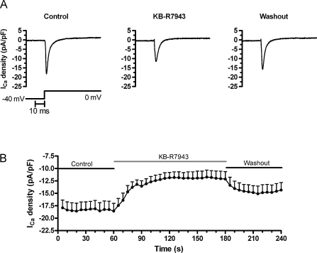
The atrioventricular node (AVN) can act as a subsidiary cardiac pacemaker if the sinoatrial node fails. In this study, we investigated the effects of the Na-Ca exchange (NCX) inhibitor KB-R7943, and inhibition of the sarcoplasmic reticulum calcium ATPase (SERCA), using thapsigargin or cyclopiazonic acid (CPA), on spontaneous action potentials (APs) and [Ca(2+)](i) transients from cells isolated from the rabbit AVN. Spontaneous [Ca(2+)](i) transients were monitored from undialysed AVN cells at 37°C using Fluo-4. In separate experiments, spontaneous APs and ionic currents were recorded using the whole-cell patch clamp technique. Rapid application of 5 μM KB-R7943 slowed or stopped spontaneous APs and [Ca(2+)](i) transients. However, in voltage clamp experiments in addition to blocking NCX current (I(NCX)) KB-R7943 partially inhibited L-type calcium current (I(Ca,L)). Rapid reduction of external [Na(+)] also abolished spontaneous activity. Inhibition of SERCA (using 2.5 μM thapsigargin or 30 μM CPA) also slowed or stopped spontaneous APs and [Ca(2+)](i) transients. Our findings are consistent with the hypothesis that sarcoplasmic reticulum (SR) Ca(2+) release influences spontaneous activity in AVN cells, and that this occurs via [Ca(2+)](i)-activated I(NCX); however, the inhibitory action of KB-R7943 on I(Ca,L) means that care is required in the interpretation of data obtained using this compound.


Figures

Fig. 1
Fig. 1
Effects of KB-R7943 on spontaneous action potentials and [Ca2+]i transients. (A) KB-R7943 inhibits spontaneous action potentials. The top trace shows a representative slow time-base recording of membrane potential from an AVN myocyte before, during and after exposure to 5 μM KB-R7943, as indicated by the horizontal bars above the trace. The lower traces show sections of the top panel from the periods indicated, in the absence (left) and presence (middle and right) of KB-R7943, displayed at a faster time-base. The time scale bar in the lower left panel applies to all three lower panels. (B) KB-R7943 inhibits spontaneous [Ca2+]i transients. The top trace shows a slow time-base averaged fluorescence plot of confocal line-scan image from an AVN myocyte (different cell from A) before, during and after application of 5 μM KB-R7943, as indicated above the trace. The lower traces show sections of the top panel from the periods indicated, in the absence (left) and presence (middle and right) of KB-R7943, displayed at a faster time-base. The time scale bar in the lower left panel applies to all three lower panels.
Fig. 2
Fig. 2
KB-R7943 inhibits the nickel-sensitive INCX in rabbit AVN cells. (Ai) Mean net current densities recorded by a ramp protocol from +60 to −80 mV (duration = 250 ms, holding potential = −40 mV, frequency = 0.33 Hz) from AVN myocytes (n = 5) during superfusion with the control solution and exposure to 5 μM KB-R7943 or 5 mM nickel chloride. (Aii) Control – nickel represents the nickel-sensitive INCX density by subtracting the current density during exposure to nickel from that in control; control – KB-R7943 represents the KB-R7943-sensitive current; and (control – nickel) − (control – KB-R7943) represents the residual nickel-sensitive INCX after KB-R7943 inhibition. *P < 0.05, **P < 0.01: control – nickel versus control – KB-R7943. (Bi and Bii) Representative time-courses of the net current densities obtained at +60 mV (Bi) and −80 mV (Bii) from an AVN cell during superfusion with the control solution and exposure to 5 μM KB-R7943 and 5 mM nickel as indicated.
Fig. 3
Fig. 3
KB-R7943 inhibits ICa,L in rabbit AVN cells. (A) Representative ICa,L traces from an AVN myocyte in the absence (control) and presence of 5 μM KB-R7943 and on washout, as indicated. The corresponding protocol used to elicit ICa,L and the time scale bar are shown underneath. (B) The time-course of mean ICa,L densities (at 0 mV, protocol shown as in A) from AVN myocytes (n = 12) in absence (control) and presence of 5 μM KB-R7943 (and following washout), as indicated.
Fig. 4
Fig. 4
Effects of thapsigargin on spontaneous action potentials and [Ca2+]i transients. (A) Thapsigargin inhibits spontaneous action potentials. The top trace shows a slow time-base recording of membrane potential from a representative AVN myocyte before, during and after application of 2.5 μM thapsigargin, as indicated above the trace. The lower traces show sections of the top panel from the periods indicated, in the absence (left) and presence (middle and right) of thapsigargin, displayed at a faster time-base. The time scale bar in the lower left trace applies to all three lower traces. In the first few seconds after thapsigargin exposure, initially the spontaneous AP rate increased and then decreased, with cessation of activity at ∼60 s after thapsigargin exposure. (B) Thapsigargin inhibits spontaneous [Ca2+]i transients. The top trace shows a slow time-base averaged fluorescence plot of a confocal line-scan image from a representative AVN myocyte (different cell from A) before, during and after application of 2.5 μM thapsigargin as indicated above the trace. The lower panels show sections of the top trace from the periods indicated, in the absence (left) and presence (middle and right) of thapsigargin, displayed at a faster time-base. The time scale bar in the lower left trace applies to all three lower traces. In the first few seconds after thapsigargin exposure, initially the [Ca2+]i transient rate increased, and the diastolic calcium baseline was elevated; the spontaneous [Ca2+]i transient rate then decreased and stopped at ∼20 s following thapsigargin exposure.

References

    1. Meijler F.L., Janse M.J. Morphology and electrophysiology of the mammalian atrioventricular node. Physiol. Rev. 1988;68:608–647. - PubMed
    1. Tawara S. Fischer; Jena, Germany: 1906. Das Reizleitungssystem des Saugetierherzens.
    1. Rigg L., Terrar D.A. Possible role of calcium release from the sarcoplasmic reticulum in pacemaking in guinea-pig sino-atrial node. Exp. Physiol. 1996;81:877–880. - PubMed
    1. Hata T., Noda T., Nishimura M., Watanabe Y. The role of Ca2+ release from sarcoplasmic reticulum in the regulation of sinoatrial node automaticity. Heart Vessels. 1996;11:234–241. - PubMed
    1. Li J., Qu J., Nathan R.D. Ionic basis of ryanodine's negative chronotropic effect on pacemaker cells isolated from the sinoatrial node. Am. J. Physiol. 1997;273:H2481–H2489. - PubMed

Publication types

MeSH terms

Substances