

Gray matter atrophy rate as a marker of disease progression in AD
- PMID: 21163551
- PMCID: PMC3657171
- DOI: 10.1016/j.neurobiolaging.2010.11.001
Gray matter atrophy rate as a marker of disease progression in AD
Abstract
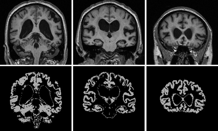
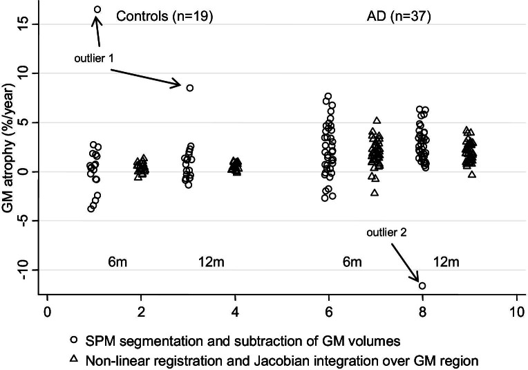
Global gray matter (GM) atrophy rates were quantified from magnetic resonance imaging (MRI) over 6- and 12-month intervals in 37 patients with Alzheimer's disease (AD) and 19 controls using: (1) nonlinear registration and integration of Jacobian values, and (2) segmentation and subtraction of serial GM volumes. Sample sizes required to power treatment trials using global GM atrophy rate as an outcome measure were estimated and compared between the 2 techniques, and to global brain atrophy measures quantified using the boundary shift integral (brain boundary shift integral; BBSI) and structural image evaluation, using normalization, of atrophy (SIENA). Increased GM atrophy rates (approximately 2% per year) were observed in patients compared with controls. Although mean atrophy rates provided by Jacobian integration were smaller than those from segmentation and subtraction of GM volumes, measurement variance was reduced. The number of patients required per treatment arm to detect a 20% reduction in GM atrophy rate over a 12-month follow-up (90% power) was 202 (95% confidence interval [CI], 118-423) using Jacobian integration and 2047 (95% CI 271 to > 10,000) using segmentation and subtraction. Comparable sample sizes for whole brain atrophy were 240 (95% CI, 142-469) using the BBSI and 196 (95% CI, 110-425) using SIENA. Jacobian integration could be useful for measuring GM atrophy rate in Alzheimer's disease as a marker of disease progression and treatment efficacy.
Copyright © 2012 Elsevier Inc. All rights reserved.
Figures
References
-
- Aljabar P., Bhatia K.K., Murgasova M., Hajnal J.V., Boardman J.P., Srinivasan L., Rutherford M.A., Dyet L.E., Edwards A.D., Rueckert D. Assessment of brain growth in early childhood using deformation-based morphometry. Neuroimage. 2008;39:348–358. - PubMed
-
- Anderson V.M., Bartlett J.W., Fox N.C., Fisniku L., Miller D.H. Detecting treatment effects on brain atrophy in relapsing remitting multiple sclerosis: sample size estimates. J. Neurol. 2007;254:1588–1594. - PubMed
-
- Anderson V.M., Fernando K.T., Davies G.R., Rashid W., Frost C., Fox N.C., Miller D.H. Cerebral atrophy measurement in clinically isolated syndromes and relapsing remitting multiple sclerosis: a comparison of registration-based methods. J. Neuroimaging. 2007;17:61–68. - PubMed
-
- Ashburner J., Friston K.J. Unified segmentation. Neuroimage. 2005;26:839–851. - PubMed
-
- Balthazar M.L., Yasuda C.L., Pereira F.R., Pedro T., Damasceno B.P., Cendes F. Differences in grey and white matter atrophy in amnestic mild cognitive impairment and mild Alzheimer's disease. Eur. J. Neurol. 2009;16:468–474. - PubMed
Publication types
MeSH terms
Grants and funding
LinkOut - more resources
Full Text Sources
Medical
