. 2011 Apr;25(4):1176-87.
doi: 10.1096/fj.10-175349. Epub 2010 Dec 16.

WNT4 is a key regulator of normal postnatal uterine development and progesterone signaling during embryo implantation and decidualization in the mouse

Affiliations

WNT4 is a key regulator of normal postnatal uterine development and progesterone signaling during embryo implantation and decidualization in the mouse

Heather L Franco et al. FASEB J. 2011 Apr.

Abstract

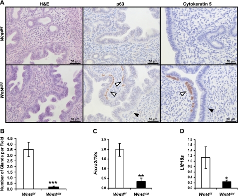
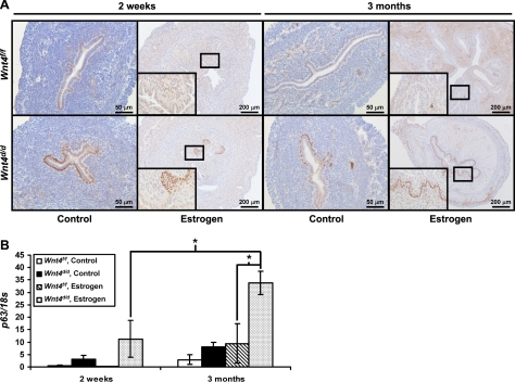
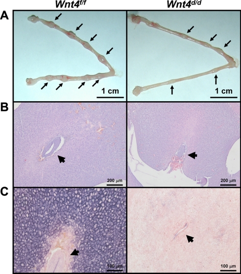
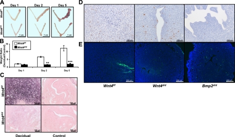
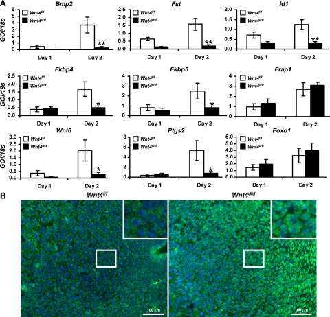
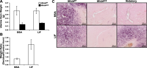
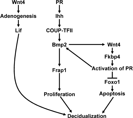
WNT4, a member of the Wnt family of ligands, is critical for the development of the female reproductive tract. Analysis of Wnt4 expression in the adult uterus during pregnancy indicates that it may play a role in the regulation of endometrial stromal cell proliferation, survival, and differentiation, which is required to support the developing embryo. To investigate the role of Wnt4 in adult uterine physiology, conditional ablation of Wnt4 using the PR(cre) mouse model was accomplished. Ablation of Wnt4 rendered female mice subfertile due to a defect in embryo implantation and subsequent defects in endometrial stromal cell survival, differentiation, and responsiveness to progesterone signaling. In addition to altered stromal cell function, the uteri of PR(cre/+)Wnt4(f/f) (Wnt4(d/d)) mice displayed altered epithelial differentiation characterized by a reduction in the number of uterine glands and the emergence of a p63-positive basal cell layer beneath the columnar luminal epithelial cells. The altered epithelial cell phenotype was further escalated by chronic estrogen treatment, which caused squamous cell metaplasia of the uterine epithelium in the Wnt4(d/d) mice. Thus, WNT4 is a critical regulator not only of proper postnatal uterine development, but also embryo implantation and decidualization.


Figures

Figure 1.
Figure 1.
Wnt4d/d mice exhibit altered postnatal uterine development. A) Analysis of 6-wk-old Wnt4f/f and Wnt4d/d mice. Left panel: hematoxylin and eosin staining. Middle panel: immunohistochemical analysis of p63. Right panel: immunohistochemical analysis of cytokeratin 5. Counterstaining is hematoxylin. Scale bars = 50 μm. White arrowheads mark p63+ cells. Black arrowheads mark p63-negative cells. B) Quantification of number of uterine glands per field in Wnt4f/f and Wnt4d/d uteri at 6 wk of age. C) Quantitative RT-PCR analysis of the gland marker Foxa2 on day −1 in the artificial decidual response. D) Quantitative RT-PCR analysis of Lif on day −1 in the artificial decidual response. Results represent means ± se; n = 5 mice/genotype. *P < 0.05; **P < 0.01; ***P < 0.001.
Figure 2.
Figure 2.
Wnt4 regulates epithelial differentiation in response to E2. A) Immunohistochemical analysis of p63 in Wnt4f/f and Wnt4d/d mice treated with vehicle or E2 for 2 wk or 3 mo. Scale bars = 50 (left panels; 200 μm (right panels). Insets: ×40 view. B) Quantitative RT-PCR analysis of p63 in Wnt4f/f and Wnt4d/d mice treated with vehicle or E2 for 2 wk or 3 mo. Results represent means ± se; n = 5 mice/group. *P < 0.05.
Figure 3.
Figure 3.
Embryo implantation is impaired in Wnt4d/d mice. A) Gross histology of Wnt4f/f and Wnt4d/d uteri on d5.5. Arrows mark implantation sites. B) Hematoxylin-and-eosin staining of d5.5 embryo implantation sites in Wnt4f/f and Wnt4d/d uteri. Arrowheads mark the embryo. C) Alkaline phosphatase staining of Wnt4f/f and Wnt4d/d implantation sites on d5.5. Positive staining is visualized as purple; counterstaining is nuclear fast red. Arrowheads mark the embryo. Scale bars = 1 cm (A); 200 μm (B); 100 μm (C).
Figure 4.
Figure 4.
Wnt4d/d mice exhibit a decidualization defect. A) Gross histology of Wnt4f/f and Wnt4d/d uteri at 1, 2, and 5 d after administration of decidual trauma in the artificial decidual response. Decidual horn is at right; control horn is at left. B) Quantification of decidual response in Wnt4f/f and Wnt4d/d mice at 1, 2, and 5 d after administration of decidual trauma in the artificial decidual response by taking the weight ratio of the decidual vs. control horn. C) Alkaline phosphatase staining of Wnt4f/f and Wnt4d/d decidual and control horns on day 2. Positive staining is visualized as purple; counterstaining is nuclear fast red. D) Immunohistochemical analysis of phospho-histone H3 in Wnt4f/f, Wnt4d/d, and Bmp2d/d uteri on day 2. Counterstaining is hematoxylin. E) TUNEL analysis of Wnt4f/f, Wnt4d/d, and Bmp2d/d uteri on day 2. Nuclei are counterstained with DAPI. Scale bars = 1 cm (A); 100 μm (C, D); 200 μm (E). Results represent means ± se; n = 5 mice/group. **P < 0.01; ***P < 0.001.
Figure 5.
Figure 5.
WNT4 regulates P4 action during decidualization. A) Quantitative RT-PCR analysis of decidual marker genes Bmp2, Fst, Id1, Fkbp4, Fkbp5, Frap1, Ptgs2, Wnt6, and Foxo1 in Wnt4f/f and Wnt4d/d uteri on days 1 and 2. B) Immunofluorescence analysis of FOXO1 in Wnt4f/f and Wnt4d/d uteri on day 2. Positive staining is visualized as green fluorescence; counterstaining is DAPI (blue). GOI, gene of interest. Scale bars = 100 μm. Results represent means ± se; n = 5 mice/group. *P < 0.05; **P < 0.01.
Figure 6.
Figure 6.
Administration of recombinant LIF is unable to rescue the Wnt4d/d decidual defect. A) Weight of uterine horns of Wnt4f/f and Wnt4d/d mice at 2 d after administration of decidual trauma in the artificial decidual response. Mice received intrauterine uterine injections of either vehicle (BSA) or LIF 1 d prior to decidual trauma; n = 5/group. B) Weight ratio of decidual vs. control horns in Wnt4f/f mice that received an injection of LIF instead of nidatory E2 at 2 d after decidual trauma in the artificial decidual response; n = 3/group. C) Alkaline phosphatase staining in Wnt4f/f and Wnt4d/d mice that received intrauterine vehicle (BSA) or LIF injections during decidualization (Wnt4f/f, Wnt4d/d) or instead of nidatory E2 (nidatory). Positive staining is visualized as purple; counterstaining is nuclear fast red. Scale bars = 100 μm. Results represent means ± se.
Figure 7.
Figure 7.
Model of WNT4 action during embryo implantation.

References

    1. Franco H. L., Jeong J. W., Tsai S. Y., Lydon J. P., DeMayo F. J. (2008) In vivo analysis of progesterone receptor action in the uterus during embryo implantation. Semin. Cell Dev. Biol. 19, 178–186 - PubMed
    1. Lee K. Y., DeMayo F. J. (2004) Animal models of implantation. Reproduction 128, 679–695 - PubMed
    1. Wang H., Dey S. K. (2006) Roadmap to embryo implantation: clues from mouse models. Nat. Rev. 7, 185–199 - PubMed
    1. Lee K. Y., Jeong J. W., Wang J., Ma L., Martin J. F., Tsai S. Y., Lydon J. P., DeMayo F. J. (2007) Bmp2 is critical for the murine uterine decidual response. Mol. Cell. Biol. 27, 5468–5478 - PMC - PubMed
    1. Li Q., Kannan A., Wang W., Demayo F. J., Taylor R. N., Bagchi M. K., Bagchi I. C. (2007) Bone morphogenetic protein 2 functions via a conserved signaling pathway involving Wnt4 to regulate uterine decidualization in the mouse and the human. J. Biol. Chem. 282, 31725–31732 - PubMed

Publication types