. 2011 Mar;120(1):42-58.
doi: 10.1093/toxsci/kfq379. Epub 2010 Dec 16.

Estrogen-like activity of perfluoroalkyl acids in vivo and interaction with human and rainbow trout estrogen receptors in vitro

Affiliations

Estrogen-like activity of perfluoroalkyl acids in vivo and interaction with human and rainbow trout estrogen receptors in vitro

Abby D Benninghoff et al. Toxicol Sci. 2011 Mar.

Abstract

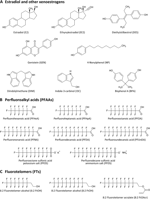
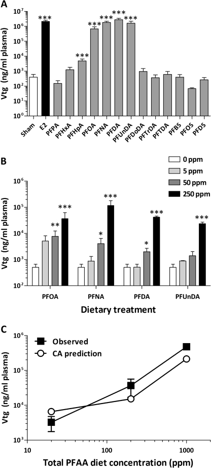
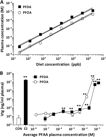
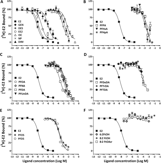
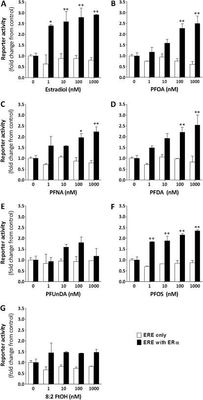
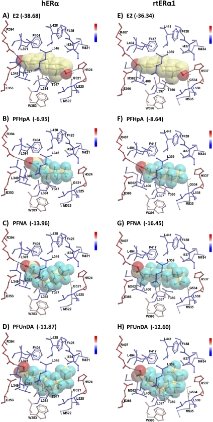
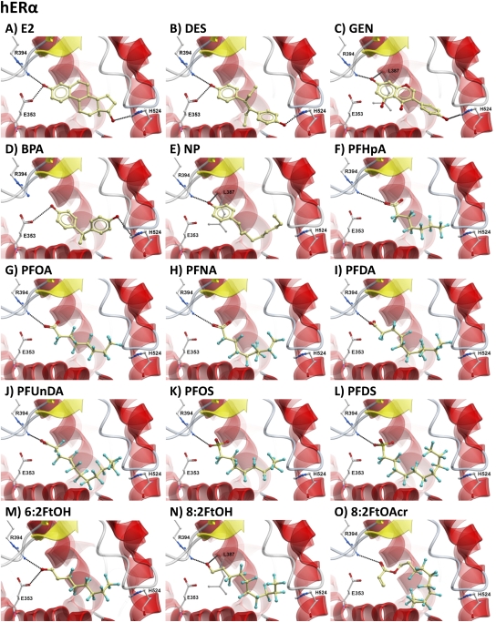
The objectives of this study were to determine the structural characteristics of perfluoroalkyl acids (PFAAs) that confer estrogen-like activity in vivo using juvenile rainbow trout (Oncorhynchus mykiss) as an animal model and to determine whether these chemicals interact directly with the estrogen receptor (ER) using in vitro and in silico species comparison approaches. Perfluorooctanoic (PFOA), perfluorononanoic (PFNA), perfluorodecanoic (PFDA), and perfluoroundecanoic (PFUnDA) acids were all potent inducers of the estrogen-responsive biomarker protein vitellogenin (Vtg) in vivo, although at fairly high dietary exposures. A structure-activity relationship for PFAAs was observed, where eight to ten fluorinated carbons and a carboxylic acid end group were optimal for maximal Vtg induction. These in vivo findings were corroborated by in vitro mechanistic assays for trout and human ER. All PFAAs tested weakly bound to trout liver ER with half maximal inhibitory concentration (IC(50)) values of 15.2-289 μM. Additionally, PFOA, PFNA, PFDA, PFUnDA, and perlfuorooctane sulfonate (PFOS) significantly enhanced human ERα-dependent transcriptional activation at concentrations ranging from 10-1000 nM. Finally, we employed an in silico computational model based upon the crystal structure for the human ERα ligand-binding domain complexed with E2 to structurally investigate binding of these putative ligands to human, mouse, and trout ERα. PFOA, PFNA, PFDA, and PFOS all efficiently docked with ERα from different species and formed a hydrogen bond at residue Arg394/398/407 (human/mouse/trout) in a manner similar to the environmental estrogens bisphenol A and nonylphenol. Overall, these data support the contention that several PFAAs are weak environmental xenoestrogens of potential concern.


Figures

FIG. 1.
FIG. 1.
Structures for known xenoestrogens (A), select PFAAs (B), and fluorotelomer (C) compounds are shown.
FIG. 2.
FIG. 2.
Effects of subchronic dietary exposure to select PFAAs and fluorotelomers on plasma Vtg concentration. (A) Mean blood plasma Vtg values are shown on a logarithmic scale + SEM (N = 18 for control, N = 12 for E2, and N = 6 for all other treatments). The diet concentration was 250 ppm (approximately 5 mg/kg bw/day) for all perfluoroalkyl compounds and 5 ppm for E2, the positive control. (B) For individual chemical treatments, fish were fed diets containing 0, 5, 50, or 250 ppm of PFOA, PFNA, PFDA, and PFUnDA. Mean blood plasma Vtg values are shown on a logarithmic scale + SEM (N = 18 for control and N = 6 for all other treatments). (C) The mixture treatments were prepared by adding equal amounts of each constituent chemical to achieve total concentrations of 20, 200, and 1000 ppm as described in the “Materials and Methods” section. Closed squares represent the measured mean blood plasma Vtg values ± SEM for the mixture treatments (N = 6). Open circles represent the predicted level of Vtg response at each concentration calculated using a simple CA model. *p < 0.05, **p < 0.01, and ***p < 0.001 compared with control (for B, within a diet group) as determined by one-way ANOVA with Dunnett's multiple comparisons post hoc test.
FIG. 3.
FIG. 3.
Dose-dependent effects of subchronic dietary exposure to PFOA and PFDA on chemical concentrations in blood plasma and Vtg expression. (A) Blood plasma concentrations of PFOA and PFDA following 2-week exposure to a broad range of diet concentrations (0.0256–2000 ppm) are shown (N = 8) in a log-log plot. Results of linear regression analyses of nontransformed data are represented by the solid lines, whereas the 95% confidence intervals are represented by the dashed lines. (B) Mean blood plasma Vtg values are shown ± SEM (N = 8) for PFOA- and PFDA-exposed fish plotted according to the measured blood concentrations of PFOA and PFDA, respectively. *p < 0.05 and **p < 0.01 compared with the 0 ppm treatment group (CON) as determined by one-way ANOVA with Dunnett's multiple comparisons post hoc test.
FIG. 4.
FIG. 4.
Binding of known xenoestrogens and various PFCs to the trout hepatic ER. Competition assays were performed with 1 mg cytosolic protein in the presence of 4nM [3H]-estradiol and increasing concentrations of test ligands, including known xenoestrogens (A), perfluoroalkyl carboxylic acids of increasing carbon chain length (B–D), perfluoroalkyl sulfonic acids (E), and fluorotelomers (F). Symbols represent mean ± SEM specific binding for each test chemical.
FIG. 5.
FIG. 5.
Transactivation of hERα by PFAAs. HEK-293T cells were transfected with (solid bar) or without (open bar) a human ERα-expressing plasmid, the XTEL luciferase reporter plasmid containing a consensus ERE sequence, and a β-galactosidase–containing plasmid for normalization of raw luminescence data. Twenty-four hours after transfection, cells were treated with increasing concentrations of E2 (A), PFOA (B), PFNA (C), PFDA (D), PFUnDA (E), PFOS (F), and 8:2FtOH (G). Values are the mean gene reporter activity expressed as fold change with respect to control (0nM treatment) (N = 3 replicate experiments). Within the ERE + ERα data set, *p < 0.05, **p < 0.01 as determined by one-way ANOVA with Bonferonni post hoc tests compared with 0nM.
FIG. 6.
FIG. 6.
Effect of fluorinated carbon chain length on perfluoroalkyl carboxylic acid docking orientation and efficiency for human or trout ERα proteins. The docked orientation of E2 is represented by the stick/space-filling model in the binding pocket of the receptor (A and E). Dockings of three representative perfluoroalkyl carboxylic acids with different fluorinated carbon chain length are shown, including PFHpA (seven carbons), PFNA (nine carbons), and PFUnDA (11 carbons), with hERα (B–D) or rtERα1 (F–H). Interacting residues of the LBDs are shown as sticks and are colored according to their hydrophobicity (inset scale) with hydrophobic residues colored blue and hydrophilic colored red. Hydrogen bonds are represented by black dotted lines between the donor (D) and the acceptor (A) and are defined as follows: Distance D–A: 2.8–3.2 Å; Angle D–H–A: 140–180°. Residue labels are numbered according to the indicated protein sequences, which are available in Supplementary figure 3. The corresponding ICM docking scores are indicated for each ligand-protein pair; scores for all PFAAs tested are available in Table 3.
FIG. 7.
FIG. 7.
In silico model showing docking of estrogens and select PFCs into the hERα LBD. Docking of test ligands into the hERα LBD was performed using Molsoft ICM, and the lowest ICM-scored poses for each calculated ligand-protein docking are shown. Ligands are colored by atom type (the carbon atoms in tan) and are displayed as ball and sticks. Relevant protein residues, including E353, R394, H524, and L387, are displayed as ball and sticks and colored by atom type: carbon atoms in gray, oxygen in red, sulfur in yellow, and fluorine in cyan. Hydrogen bonds are represented by black dotted lines between the donor (D) and the acceptor (A) and are defined as follows: Distance D–A: 2.8–3.2 Å; Angle D–H–A: 140–180°.

References

    1. Abagyan R, Totrov M, Kuznetsov D. ICM—a new method for protein modeling and design: applications to docking and structure prediction from the distorted native conformation. J. Comput. Chem. 1994;15:488–506.
    1. Abdellatif AG, Preat V, Taper HS, Roberfroid M. The modulation of rat liver carcinogenesis by perfluorooctanoic acid, a peroxisome proliferator. Toxicol. Appl. Pharmacol. 1991;111:530–537. - PubMed
    1. Benninghoff AD, Williams DE. Identification of a transcriptional fingerprint of estrogen exposure in rainbow trout liver. Toxicol. Sci. 2008;101:65–80. - PMC - PubMed
    1. Bisson WH, Koch DC, O'Donnell EF, Khalil SM, Kerkvliet NI, Tanguay RL, Abagyan R, Kolluri SK. Modeling of the aryl hydrocarbon receptor (AhR) ligand binding domain and its utility in virtual ligand screening to predict new AhR ligands. J. Med. Chem. 2009;52:5635–5641. - PMC - PubMed
    1. Blair RM, Fang H, Branham WS, Hass BS, Dial SL, Moland CL, Tong W, Shi L, Perkins R, Sheehan DM. The estrogen receptor relative binding affinities of 188 natural and xenochemicals: structural diversity of ligands. Toxicol. Sci. 2000;54:138–153. - PubMed

Publication types

MeSH terms