. 2011 Feb 24;117(8):2396-404.
doi: 10.1182/blood-2010-04-278788. Epub 2010 Dec 16.

A mechanistic rationale for MEK inhibitor therapy in myeloma based on blockade of MAF oncogene expression

Affiliations

A mechanistic rationale for MEK inhibitor therapy in myeloma based on blockade of MAF oncogene expression

Christina M Annunziata et al. Blood. .

Abstract

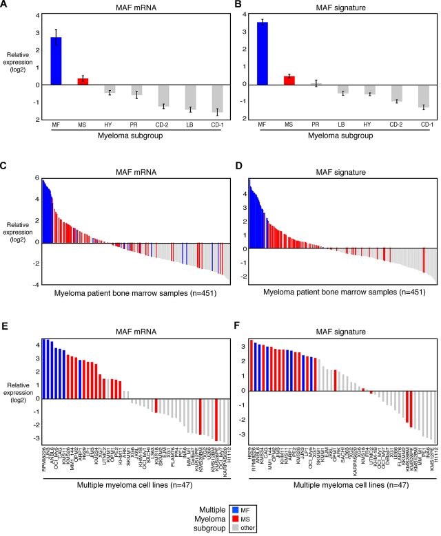
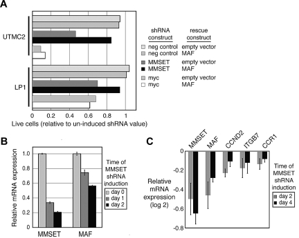
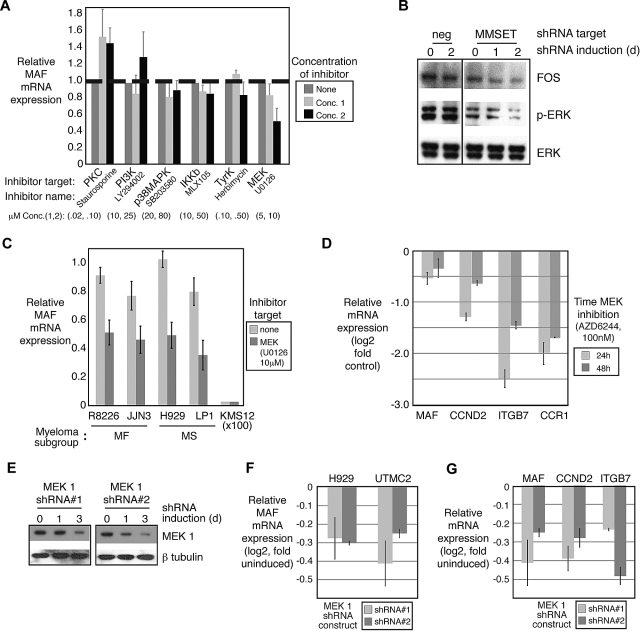
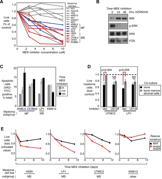
Modulating aberrant transcription of oncogenes is a relatively unexplored opportunity in cancer therapeutics. In approximately 10% of multiple myelomas, the initiating oncogenic event is translocation of musculoaponeurotic fibrosarcoma oncogene homolog (MAF), a transcriptional activator of key target genes, including cyclinD2. Our prior work showed that MAF is up-regulated in an additional 30% of multiple myeloma cases. The present study describes a common mechanism inducing MAF transcription in both instances. The second mode of MAF transcription occurred in myelomas with multiple myeloma SET domain (MMSET) translocation. MMSET knockdown decreased MAF transcription and cell viability. A small-molecule screen found an inhibitor of mitogen-activated protein kinase kinase (MEK), which activates extracellular signal-regulated kinase (ERK)-MAP kinases, reduced MAF mRNA in cells representing MMSET or MAF subgroups. ERK activates transcription of FOS, part of the AP-1 transcription factor. By chromatin immunoprecipitation, FOS bound the MAF promoter, and MEK inhibition decreased this interaction. MEK inhibition selectively induced apoptosis in MAF-expressing myelomas, and FOS inactivation was similarly toxic. Reexpression of MAF rescued cells from death induced by MMSET depletion, MEK inhibition, or FOS inactivation. The data presented herein demonstrate that the MEK-ERK pathway regulates MAF transcription, providing molecular rationale for clinical evaluation of MEK inhibitors in MAF-expressing myeloma.


Figures

Figure 1
Figure 1
MAF and target genes are highly expressed in myelomas with MAF or MMSET translocations. Expression of MAF and target genes in primary MM patient samples were derived from Affymetrix U133plus2.0 gene expression profiling data from 451 purified bone marrow plasma cell populations collected from untreated patients with MM (A-D) or 47 MM cell lines (E-F). Samples are ranked according to the expression of MAF itself (A,C,E, average of 4 probe sets) or the average expression of MAF and 3 target genes (B,D,F). Expression was centered based on the mean value in normal plasma cells (GEO dataset GSE5900). Myeloma subgroups are designated according to Zhan et al.
Figure 2
Figure 2
MMSET regulates MAF transcription. (A) Myeloma cell lines UTMC2 and LP-1, both harboring the MMSET translocation, either were engineered to express MAF cDNA under a constitutive LTR promoter or contained an empty construct. These cells were secondarily transduced with a retrovirus expressing MMSET shRNA and coexpressing GFP (“Retrovirally mediated transduction”). Live, GFP-expressing cells were quantified by flow cytometry and normalized to day 2 after retroviral transduction. (B) MM cells were transduced with a TET-inducible MMSET shRNA construct and selected. MMSET shRNA was induced for the indicated times, and RNA was quantified by real-time PCR. (C) Gene expression was profiled during inducible MMSET knockdown. Experiments were repeated in triplicate. Shown are average values of target genes compared with control shRNA targeting luciferase (± SEM).
Figure 3
Figure 3
MEK signaling regulates MAF transcription. (A) Myeloma cell line LP1 was treated with small-molecule kinase inhibitors targeting major cell signaling cascades at the indicated concentrations. MAF mRNA was measured by quantitative RT-PCR. (B) LP1 MM cells were transduced with a TET-inducible MMSET shRNA construct, selected, and MMSET shRNA was induced for the indicated times. Protein lysates were analyzed by Western blot. ERK phosphorylation and FOS expression were decreased in these cells. (C) MAF-expressing myeloma cell lines were treated with MEK inhibitor U0126 (10μM), and MAF mRNA was measured by real-time quantitative PCR. (D) MAF and target gene mRNA were measured by quantitative RT-PCR after treatment with a second MEK inhibitor, AZD6244 100nM, for the indicated times. (E) MM cells transduced with 2 individual TET-inducible MEK1 shRNA construct, selected, and MEK1 shRNA was induced for the indicated times. Protein lysates were analyzed by Western blot to confirm MEK1 protein decrease after shRNA-mediated knockdown. (F) MAF expression was quantified by quantitative RT-PCR after MEK1 depletion with each shRNA construct in 2 MM cell lines. (G) MAF and target gene mRNA expression were measured by quantitative RT-PCR after MEK1 depletion. Error bars represent mean plus or minus SD.
Figure 4
Figure 4
MEK inhibitor toxicity is higher in MAF-expressing MM cell lines and rescued by MAF reexpression. (A) MM cell lines were incubated with indicated concentrations of MEK inhibitor U0126, and cell number was enumerated by flow cytometry after 8 days. Live cells were normalized to solvent (dimethyl sulfoxide)–treated control. (B) Proapoptotic BIM is up-regulated after MEK inhibition. Western blot also shows decreased ERK phosphorylation and FOS expression. (C) MEK inhibitor-induced apoptosis was detected by VAD-FITC labeling of cleaved caspase 3 in MM cell lines treated with U0126 (10μM) at the indicated time points. (D) MM cell lines were incubated with either dexamethasone (24nM) or U0126 (10μM) for 7 days in the presence or absence of HS-5 bone marrow stromal cell line. Live cells were quantified by sodium 3-[1-[(phenylamino) carbonyl]-2H-tetrazolium hydroxide assay and normalized to untreated control cells (± SEM). (E) MAF transgene was stably and constitutively expressed in MM cell lines using an exogenous LTR promoter. Live cells were enumerated by flow cytometry for up to 11 days of MEK inhibition (U0126, 10μM). Shown are representative growth curves from at least 3 independent experiments.
Figure 5
Figure 5
FOS regulates MAF transcription. (A) Chromatin immunoprecipitation of FOS demonstrates binding to MAF promoter in MM cells that is decreased with MEK inhibition (U0126, 10μM). (B) MEK inhibition (U0126, 10μM) decreased FOS binding to MAF locus by chromatin immunoprecipitation in MAF-expressing cell lines. (C) Knockdown of FOS is toxic to MAF-overexpressing myeloma cell lines. The indicated myeloma cell lines were transduced with a retrovirus expressing FOS shRNA and coexpressing GFP (“Retrovirally mediated transduction”). Live GFP+ cells were enumerated by fluorescence-activated cell sorter and normalized to the value at day 2 after retroviral infection. Reexpression of MAF rescues myeloma cells transduced with an shRNA targeting FOS. (D) Quantitative RT-PCR measurement of FOS mRNA levels after induction of the FOS shRNA in H929 cells. (E) Western blot of FOS protein levels after introduction of the FOS shRNA in H929 cells.

Comment in

  • MEK and MAF in myeloma therapy.
    Popovic R, Licht JD. Popovic R, et al. Blood. 2011 Feb 24;117(8):2300-2. doi: 10.1182/blood-2011-01-327262. Blood. 2011. PMID: 21350059

References

    1. Jemal A, Siegel R, Ward E, Murray T, Xu J, Thun MJ. Cancer statistics, 2009. CA Cancer J Clin. 2007;59(4):225–249. - PubMed
    1. Bensinger W. Stem-cell transplantation for multiple myeloma in the era of novel drugs. J Clin Oncol. 2008;26(3):480–492. - PubMed
    1. Zhan F, Huang Y, Colla S, et al. The molecular classification of multiple myeloma. Blood. 2006;108(6):2020–2028. - PMC - PubMed
    1. Eychene A, Rocques N, Pouponnot C. A new MAFia in cancer. Nat Rev Cancer. 2008;8(9):683–693. - PubMed
    1. Hurt EM, Wiestner A, Rosenwald A, et al. Overexpression of c-maf is a frequent oncogenic event in multiple myeloma that promotes proliferation and pathological interactions with bone marrow stroma. Cancer Cell. 2004;5(2):191–199. - PubMed

Publication types

MeSH terms

Substances